Home Blog Page 5

Ulcerated necrobiosis lipoidica: what is it and how should it be managed?

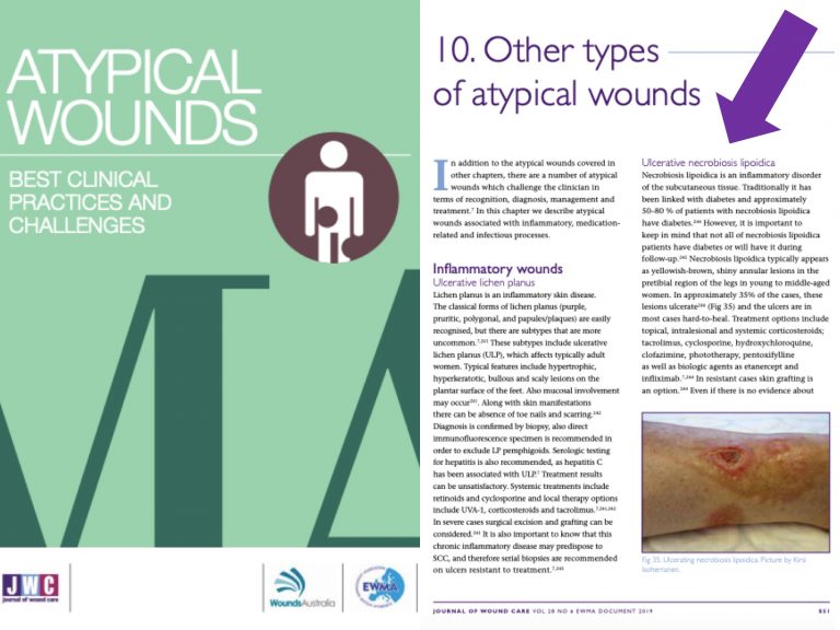
Necrobiosis lipoidica is a condition that we will always have to include if we list the causes of “atypical” leg ulcers. In fact, in the EWMA document “Atypical Wounds”,which has recently been published, this entity has its section. It is not a frequent type of wound, but it is important to know it.

What is necrobiosis lipoidica?

Necrobiosis lipoidica is a granulomatous disease of unknown cause, in which degeneration of dermal collagen occurs.

The name “necrobiosis” is due to the type of alteration found when a biopsy is performed (necrobiosis = degenerated collagen). The term “lipoidica” refers to the typical yellowish colour of the lesions.

It is a rare condition, which usually develops at middle age (30-40 years). Its incidence is higher in women. Its clinical course is usually chronic.

Although it has traditionally been associated with diabetes, mainly type I, and was once called “necrobiosis lipoidica diabeticorum, it seems that this relationship is not as strong as previously thought. In fact, although 50-80% of patients with necrobiosis lipoidica have diabetes, the incidence of necrobiosis lipoidica in diabetic patients is only 0.3-1.2%. In addition to showing up in healthy individuals, it has been described in patients with autoimmune thyroid disease, inflammatory bowel disease, or rheumatoid arthritis. However, the relationship between these pathologies and necrobiosis lipoidica is not clear and its association may be coincidental.

Its true cause and pathological mechanism are not very clear, but several theories have been proposed. The most accepted etiopathogeny is that collagen degeneration is due to alterations at the vascular level, by the deposit of immunocomplexes or occlusive microangiopathy. It has also been proposed that the formation of granulomas may involve a defect in the migration of neutrophils.

What about its clinical presentation and diagnosis?

Lesions usually occur in the lower extremities, especially in the pretibial region. However, specific cases have been described in other locations such as the head, trunk, genitals or upper extremities.

Although clinical presentation is quite characteristic, the appearance of the lesions varies according to the evolutionary phase of the process.

They begin as asymptomatic papules and nodules that become well-defined, yellow-brownish, with erythemato-violaceous edges and atrophic center, in which telangiectasias can be visualized.

Trauma can worsen lesions, as in pyoderma gangrenosum (patergia phenomenon).

Ulceration may occur in about one-third of cases. It should be borne in mind that chronic venous insufficiency, especially phlebolinfedema, can complicate the lesions, with greater extension and ulceration, in addition to making diagnosis difficult (ochre dermatitis may be clinically similar).

A biopsy will give us the diagnostic key. Necrobiosis lipoidica is histologically characterized by palisaded granulomas (aggregates of histiocytes and giant cells) arranged horizontally alternating with bands of degenerated collagen (necrobiosis). The wall of the dermal vessels may be thickened. The accompanying inflammatory infiltrate varies according to the time of evolution of the lesions. Initially the neutrophilic infiltrate is characteristic, which decreases when the atrophy develops.

How should necrobiosis lipoidica be treated?

The treatment of this condition is a real challenge. As there are no clinical trials to evaluate the real usefulness of the different treatments used in necrobiosis lipoidica, there are no established lines of treatment. Given that in some patients remission may be spontaneous, it is difficult to assess whether, in the published case series, the response is really due to the indicated treatment.

In diabetic patients, adequate diabetes control is essential.

The different treatment alternatives found in the literature are proposed with the potential objective of controlling granuloma formation, decreasing inflammation and/or favouring microcirculation.

The most commonly used treatment is topical or intralesional corticosteroids, but the use of systemic corticoids, tacrolimus, pentoxifylline, phototherapy, fumaric acid esters, cyclosporine, antimalarials, biological drugs (infliximab, etanercept) is also described… In summary, a wide range of possibilities and little evidence available… In addition, many cases are resistant to the different treatments used.

What treatments can be used for ulcerated lesions?

The goal of treatment will be the same as with non-ulcerated lesions, i.e. we will also try to control the potentially triggering mechanisms. However, a strategy to promote granulation and epithelialisation of wounds should also be developed. The therapeutic challenge will therefore be even greater.

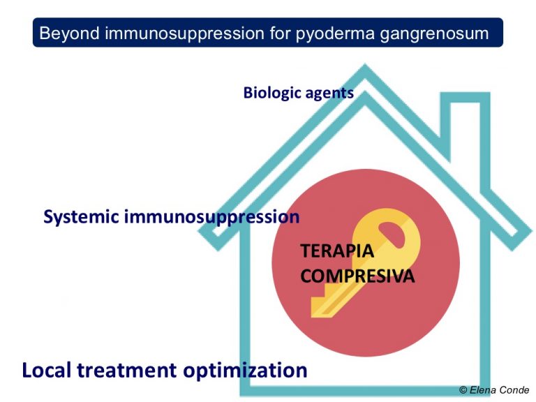
Compression therapy, as in any other leg ulcer, if there is no contraindication (always palpate pulses and perform  ABI if necessary), will help healing by its anti-gravity and anti-inflammatory effect (See post Compression is key in the treatment of leg ulcers). Once the lesions have resolved, the use of compression stockings can help in the prevention of new outbreaks.

On a local level, as in any other type of wound, we will regulate the frequency of dressings and select the dressings according to the characteristics of the wound bed and edges. Skin grafting is interesting in resistant cases.

It is interesting to remember that once a correct diagnosis has been made and adequate etiological treatment has been given, which in the case of necrobiosis lipoidica is not clear, local wound care will be characterized by similar strategies to promote an anti-inflammatory environment.

What is our experience in our clinical practice?

Our experience is very limited as it is a rare type of wound. However, we have just published the excellent result, both in pain control and wound epithelization, of the combined use of topical sevoflurane (see post: Have you ever heard of the topical use of sevoflurane in wounds?) and punch grafting, in a young patient with a very painful and progressively growing ulcer. Of course, with adjuvant compressive therapy… Another alternative for treatment! ?https://www.ncbi.nlm.nih.gov/pubmed/31412426

2491483_1185


References:

  1. Isoherranen K, O’Brien JJ, Barker J, Dissemond J, Hafner J, Jemec GBE, Kamarachev J, Läuchli S, Montero EC, Nobbe S, Sunderkötter C, Velasco ML.Atypical wounds. Best clinical practice and challenges. J Wound Care. 2019 Jun 1;28(Sup6):S1-S92. doi: 10.12968/jowc.2019.28.Sup6.S1. 
  2. Sibbald C, Reid S, Alavi A. Necrobiosis Lipoidica. Dermatol Clin. 2015 Jul;33(3):343-60. 
  3. Marcoval J, Gómez-Armayones S, Valentí-Medina F, Bonfill-Ortí M, Martínez-Molina L. Necrobiosis lipoidica: a descriptive study of 35 cases. Actas Dermosifiliogr. 2015 Jun;106(5):402-7. 

How to promote healing in ulcers secondary to pyoderma gangrenosum?

If I had to choose a single sentence to answer this question, it would undoubtedly be “with an adequate anti-inflammatory strategy and, if necessary, with an invasive approach”.

Some of you may have been surprised when Reading the term “invasive” associated with the word “gangrenous pyoderma”. In fact, the message we are used to is “pyoderma gangrenosum = no surgical debridement”.

On the other hand, many of you will think that I am not saying anything new when talking about an anti-inflammatory strategy, because treatment with systemic and topical corticosteroids is the first choice in these patients (See post: Pyoderma gangrenosum: a diagnostic and therapeutic challenge).

The aim of this post is to explain and underline the interest of a holistic approach to the patient with an ulcer secondary to pyoderma gangrenosum, beyond the use of immunosuppressive agents.

And we will explain it in 5 key points:

1. Compression Therapy

As we explained in the entry “Compression is key to treating leg wounds”, the inflammatory process inherent to any leg ulcer involves an alteration in microcirculation, with greater capillary filtration, and, due to the effect of gravity, an increase in intravenous pressure. Therefore, despite the absence of reflux or an obstructive disorder, the large inflammation found in pyoderma gangrenosum can trigger a state of venous hypertension that may hinder healing.

The interest of compression therapy in any leg injury is due to the following effects:

  • Decreased capillary filtration
  • Increased local lymphatic drainage
  • Reduction of inflammation
  • Increased arterial flow

It is important to remember that the patient with an ulcer secondary to pyoderma gangrenosum (as with any other leg ulcer) may also have venous insufficiency or peripheral artery disease.  In fact, in patients with pyoderma gangrenosum and chronic venous insufficiency, clinical worsening in spite of an adequate immunosuppressive treatment may be due to the pro-inflammatory effect of basal venous hypertension.  As in the evaluation of any leg injury, pulse palpation is obligatory. In case of doubt, we will measure the ankle-brachial index (ABI).

So, which degree of compression would be necessary?

As underlined in the chapter on Pyoderma Gangrenosum in the EWMA’s document Atypical Wounds,1 there are no studies considering this topic. However, since the application of the compression bandage may be painful initially, the recommendation is to start with 20 mmHg and increase the compression depending on patient’s tolerance. With progressive reduction of inflammation, tolerance will increase. If the patient has concomitant artery disease, the type of compression will be adapted to it (see post “Compression is key to treating leg wounds”)

2. Promotion of a suitable wound bed microenvironment, wound edges and perilesional skin

As with any chronic ulcer, the choice of dressing type and frequency of dressing changes will depend on the characteristics of the wound bed and perilesional skin. Topical corticosteroids, like the systemic corticosteroids, is the etiological treatment of choice for pyoderma gangrenosum. Topical calcineurin inhibitors (tacrolimus) are an effective and safe alternative.

A holistic evaluation has to be carried out during each dressing change in order to detect and treat at an early stage the possible cause of clinical worsening. If there is more pain, extension of the lesion and perilesional erythema, in addition to thinking of a bad response to the treatment, the possibility of infection must always be assessed (the risk of infection is increased in these patients due to immunosuppressive treatment).

3. Negative pressure therapy and skin grafts

Complete healing rates with classical immunosuppressive treatments (predominantly oral corticosteroids and cyclosporine) do not reach 50% at 6 months. Although different biologic agents may be an effective alternative in non-responders, the problem is not only controlling pyoderma activity, but also repairing all lost tissue.

Once the inflammatory activity is controlled, the strategy to facilitate the formation of granulation tissue and promote epithelialization will be similar to that used in wounds of other aetiologies. Therefore, negative pressure therapy and thin split-thickness grafts will be good alternatives, either alone or in combination, to accelerate healing. This benefit is described in cases and small series published in the literature.2,3 In our clinical practice, as you can imagine, the type of  skin graft that we normally use in these cases is punch grafting.

4. Topical Sevoflurane

Analgesic control is a key aspect in the treatment of wounds secondary to pyoderma ganrenosum, which are very painful lesions. Topical irrigation of sevoflurane is a very interesting option in these patients. Furthermore, as we commented in the post Have you ever heard of the topical use of sevoflurane in wounds?, this halogenated derivative of ether would not only have a beneficial analgesic effect, but it also has anti-inflammatory and healing properties (due to its vasodilator action).

La combinación secuencial de sevoflurano, terapia de presión negativa ambulatoria e injertos en sello es una constante en nuestra consulta.

5. Is it really a pyoderma gangrenosum?

Whenever an ulcer diagnosed as pyoderma gangrenosum does not respond to an adequate immunosuppressive treatment, besides considering that  the ulcer may be resistant to treatment and could require association or change to other drugs, we should always wonder if “is it really an ulcer secondary to pyoderma gangrenosum?”

In fact, more than 10% of ulcers diagnosed as pyoderma gangrenosum are really other types of lesions4 … Among them, for example, vasculitis, Martorell ulcer, antiphospholipid syndrome and even factitious ulcers, which are also a diagnosis of exclusion (see post “Factitial ulcer: a silent cry for help“)

In order to stop considering pyoderma gangrenosum as a diagnosis of exclusion, different expert consensus on possible diagnostic criteria have been published. The most recent proposal includes, as we can see in the table, 1 major criterion (neutrophilic infiltrate in the biopsy) and the presence of 4 of the 8 minor criteria, with a sensitivity and specificity of 86% and 90%, respectively.5

I would like to end this post with my most sincere gratitude to the organizers and participants of the VI Congreso Nacional y V Internacional de Atención al Paciente con Heridas for the enormous affection, kindness and hospitality with which they have welcomed me in Medellin. IT HAS BEEN A FANTASTIC EXPERIENCE!


Referencias:

1.  Isoherranen K, O’Brien JJ, Barker J, Dissemond J, Hafner J, Jemec GBE, Kamarachev J, Läuchli S, Montero EC, Nobbe S, Sunderkötter C, Velasco ML. Atypical wounds. Best clinical practice and challenges. J Wound Care. 2019 Jun 1;28(Sup6):S1-S92.

2.  Pichler M, Larcher L, Holzer M, Exler G, Thuile T, Gatscher B et al. Surgical treatment of pyoderma gangrenosum with negative pressure wound therapy and split thickness skin grafting under adequate immunosuppression is a valuable treatment option: Case series of 15 patients. J Am Acad Dermatol.2016 Apr;74(4):760-5.

3.  Pichler M, Thuile T, Gatscher B, Tappeiner L, Deluca J, Larcher L, et al. Systematic review of surgical treatment of pyoderma gangrenosum with negative pressure wound therapy or skin grafting. J Eur Acad Dermatol Venereol. 2017 Feb;31(2):e61-e67.

  1. Weenig RH, Davis MD, Dahl PR, Su WP. Skin ulcers misdiagnosed as pyoderma gangrenosum. N Engl J Med. 2002 Oct 31;347(18):1412-8.
  2. Maverakis E, Ma C, Shinkai K, Fiorentino D, Callen JP, Wollina U, et al. Diagnostic Criteria of Ulcerative Pyoderma Gangrenosum: A Delphi Consensus of International Experts. JAMA Dermatol.2018 Apr 1;154(4):461-466. 


Phlebolymphedema, a term that should be more used

In  our daily life as a health care professional treating patients with leg ulcers, have you ever used or heard the word PHLEBOLYMPHEDEMA?  Probably rarely or never… But you have certainly seen and treated patients with legs very similar to the ones you can see below:

What name do you use then to refer to the aetiology of these very frequent skin changes in the legs? I’m sure the range of answers is very wide, but the words LYMPHEDEMA or CHRONIC VENOUS INSUFFICIENCY will predominate. The use of this varied terminology clearly indicates that we do not call PHLEBOLYMPHEDEMA by its name.

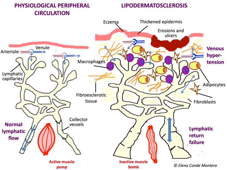
Why don´t we usually use this term, when it is the most frequent cause of lymphedema in our environment and it is very common in our consultations? The reason is undoubtedly a lack of knowledge of physiopathology. We are not clear about what is happening in the microcirculation of those swollen legs because our mind has not internalized that uncontrolled venous hypertension will end up generating a collapse and damage of the lymphatic capillaries and that, therefore, the venous edema will become venous and lymphatic.  In fact, that edema, with episodes of lesser or greater redness and progressive appearance of woody consistency, warty lesions, erosions or ulcers, and possibility of superinfection, is the result of venous insufficiency and secondary lymphatic insufficiency.

In addition, possible co-morbidities of these patients, such as congestive heart failure, cirrhosis or nephropathy, may obstruct lymphatic drainage of interstitial fluid and macromolecules.

Most of our patients with phlebolymphedema have mobilization problems, especially osteoarticular ones. This lack of activation of the muscle pump not only influences the venous return, but also the lymphatic return, since the walls of the lymphatics also need muscle contraction to overcome gravity.

Therefore, although at an academic level it is simpler to explain and study the causes of leg edema in separated compartments, in clinical practice we very often find combined aetiologies. The detection of all the factors that may be involved in the edema development is fundamental to carry out an adequate and early treatment. I want to emphasize “early” because uncontrolled edema, with the presence of proteins and other molecules in the interstitium, produces a pro-inflammatory environment that produces fibrosis in the subcutaneous cellular tissue and the vessel wall, with irreversible damage, in addition to promoting bacterial proliferation with cellulitis risk. In order to stop this lipodermatosclerosis as soon as possible, it is essential to quickly reduce the edema and maintain it over time.

Undoubtedly, using terms that take into account the different aetiologies involved, such as PHLEBOLYMPHEDEMA, means that we are very aware from the outset of the targets of our treatment.

The diagnosis of phlebolymphedema is clinical (with great importance of anamnesis), but we can also help us with imaging tests such as echo-Doppler and lymphoscintigraphy. It is characterized by signs of chronic venous insufficiency, such as phlebectasic crown in the foot or ochre coloration secondary to hemosiderin deposits, signs of lymphatic insufficiency, such as edema on the back of the feet and toes, and changes in epidermis, dermis and subcutaneous cellular tissue associated with chronic edema, such as eczema, vesicles or warty lesions (variability depending on chronicity), and the fibrous consistency typical of lipodermatosclerosis.

It is often bilateral, but usually one leg is more affected than the other. Note: a frequent differential diagnosis presented by these legs is cellulitis. However, even though the risk of infection in these patients is higher, the development of cellulitis would be only in one leg. So, even though these legs sometimes appear very red, swollen and hot… We would never suspect a bilateral cellulitis!

The idea of talking about phlebolymphedema just came to me in Zurich, at the annual meeting of the European Venous Forum. At this congress, the relevance of the alteration of the microcirculation of the lymphatic capillaries associated with venous hypertension was repeatedly discussed. Consequently, I thought about the scarce use the word PHLEBOLYMPHEDEMA, when it is the most appropriate term in many of our patients. In this meeting I also thought about another term: LIPEDEMA. I totally agree with the experts who, taking into account that the presence of edema in lipedema has not been demonstrated, propose that it would be more purist to talk about PAINFUL LIPOPHYPERTROPHY. But since the term is established, we will probably continue to talk about LIPEDEMA… So we will continue to talk inadequately…

The “wrongly named” lipedema sometimes also overlaps with phlebolymphedema and we would talk about PHLEBOLIPOLYMPHEDEMA. Furthermore, even if the typical clinical characteristics of lipedema are not met, it is not uncommon to find fat accumulation in the legs of these patients. This is because the chronic inflammation associated with phlebolymphedema not only produces fibrosis, but also stimulates the proliferation of adipocytesA perfect vicious circle that must be cut as soon as possible, because in addition to facilitating ulcers, it also increases the risk of infection.

And regarding infections, it should also be noted that the proper functioning of the lymphatic system is an essential pillar of the immune system. Each episode of cellulitis in these legs with phlebolymphedema produces irreparable damage to the lymphatics, with the consequent development of recurrent infections.

CONCLUSION: there are many reasons that support the need for rapid treatment aimed at controlling venous and lymphatic insufficiency. In this management it is very important that different professionals collaborate, since the patient is going to benefit enormously from a multidisciplinary management in which the following list should be considered:

  1. Skin hydratation and adequate hygiene

We must recommend cleaning and drying avoiding aggressive rubbing on the epidermis and that emollients are used daily. Patients must be informed of the risk of cellulitis, so they can consult if alarm signs appear (erythema, increased pain, fever). Multiple erosions and superficial ulcers in the context of hypodermitis respond very well to treatment with topical steroids and optimization of compressive therapy. The application of zinc oxide products will have a beneficial anti-inflammatory effect in the presence or not of cutaneous wounds in these legs (see post: “Why do we use topical zinc on wounds and perilesional skin?”) In case of developing ulcers of greater extension and depth, besides optimizing the local treatment, it is essential to ensure that the etiological treatment is adequate (mainly compression therapy).

  1. Change of daily habits

We must emphasize compliance with anti-edema measures and provide professional help to achieve weight loss.

  1. Sclerotherapy or other endovenous techniques

These techniques will be performed if, after an adequate ultrasound study, the detected reflux could benefit from this treatment (see post: “Sclerotherapy in venous ulcers: the sooner the better”). Unlike surgery, these are safe techniques to avoid damage to the lymphatic vessels. This correction of venous reflux, and therefore of venous hypertension, will have a direct impact on lymphatic insufficiency, which can be greatly improved.

  1. Therapeutic compression

Bandages with a high static stiffness index (see post: “Band and bandage, not the same thing”) are the most indicated in these patients to try to reduce edema as soon as possible. After an initial intensive treatment with bandages, until the greatest possible reduction in the leg perimeters is achieved, maintenance treatment with compression stockings is usually recommended. The more advanced the phlebolymphedema, it will be preferable to use flat-knit stockings, since they have less stretching capacity than circular tissue and are custom-made, which makes them safer and more effective in these patients. Since the recommended pressure in these patients is high (30-40 mmHg) and they have difficulty putting on the stockings, self-adjustable velcro wraps are an effective alternative, easy to put on, and very well accepted by patients, families and caregivers.

  1. Manual lymphatic drainage

With the hands, through very soft and slow movements, you can stimulate the lymphatic vessels to help them open and promote the drainage of the fluid from the areas with more congestion towards the proximal functional lymphatics

  1. Physical exercise

Exercises that stimulate the calf and plantar muscle pump, which facilitate venous and lymphatic return, and therefore decrease venous hypertension and improve distal perfusion, are essential. The simple dorsiflexion movement of the foot and ankle is beneficial in those patients with reduced mobility. But if there is any “specially beneficial activity” that we should recommend without doubt is aquagym. Aquatic exercises are beneficial, not only because of the movement performed, but also because of the cold and pressure exerted, with an analgesic effect. ?


References:

  1. Farrow W. Phlebolymphedema-a common underdiagnosed and undertreated problem in the wound care clinic. J Am Col Certif Wound Spec. 2010 Apr 22;2(1):14-23. 
  2. Bunke N, Brown K, Bergan J. Phlebolymphemeda: usually unrecognized, often poorly treated. Perspect Vasc Surg Endovasc Ther. 2009 Jun;21(2):65-8.


Timolol for chronic wounds?

The idea of talking about topical timolol in non-healing wounds came to my mind during the recent World Congress of Dermatology 

Before continuing, I would like to thank Marco Romanelli for the invitation to participate as a speaker in the only session about wound healing at the congress. It was a real honour to meet great experts whom I admire. I learned from their presentations and enjoyed a lot talking about the treatment Pearl in our clinic,punch grafting. As Patricia Senet and José Contreras highlighted, the figure of the dermatologist is very important in wound management, especially in wounds secondary to less frequent aetiologies (tumours, inflammation, arteriolopathy). In addition, as Patricia Senet pointed out, when faced with an ulcer that does not close, we must always ask ourselves three questions:

  1. Is the etiological treatment adequate?
  2. Is there associated arteriopathy?
  3. IS IT REALLY A VENOUS ULCER?

The dermatologist is key in this differential diagnosis 🙂

Coming back to the topic of post, let’s understand why there is growing interest in the use of topical timolol in wounds, especially among dermatologists.

What is timolol?

Timolol maleate is a non-selective beta-blocker used for the treatment of glaucoma in ophthalmology at a concentration of 0.25 or 0.5%, in solution or gel. In dermatology, it is increasingly used in the treatment of certain infantile hemangiomas, with an excellent efficacy and safety profile. Like the rest of the drugs in the beta-blocker family, it prevents the binding of catecholamines (adrenaline and noradrenaline) to the β-adrenergic receptors on the cell surface, thus inhibiting their action.

Multiple cells in the body have this type of receptors. In fact, since the stimulation of the β-adrenergic receptors of cardiac cells increases heart rate and contractility, as well as blood pressure, the use of systemic beta-blocker drugs is widespread in cardiology.

Timolol is called “non-selective” because, unlike selective beta-blockers (also called cardioselectives), it does not have a preference for a particular type of receptor.

Why does it have a potential interest in wound healing?

First of all, I have to say that I had the following mental association: non-selective beta-blockers decrease peripheral tissue perfusiónè avoid in patients with arteriosclerosis ulcers and arteriolosclerosis (like Martorell ulcer)! And in fact, this recommendation is published and there is consensus among experts.

Therefore, the first time I saw a publication about thymolol in wounds, it caught my attention. Then, by digging deeper into the real impact of beta-blockers on peripheral skin microcirculation, there seems to be controversy.1

But well, analyzing in more detail the relationship of systemic beta-blockers with wound healing, in burns it has been found benefit of propanolol to decrease the damage caused by the typical hypermetabolic response in these patients, shortening and improving the healing process.2

What actions are associated with timolol in order to understand its potential beneficial effect on healing?

The different cells involved in healing have beta-adrenergic receptors.

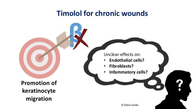
The interest in topical timolol arises from the in vitro finding of its stimulating effect on keratinocyte migration. The activation of the β-adrenergic receptors of the keratinocyte membrane slows down the cascade of signals involved in their migration and modifies cellular polarization and response to electrical stimuli, which is fundamental for their migration. Therefore, blocking these receptors would be positive in favour of keratinocyte migration.3


This beneficial effect on cell migration seems to occur also in endothelial cells. The migration of endothelial cells stimulates endothelial growth factor, with the consequent increase in angiogenesis. This increase in angiogenesis has been evidenced in studies with rats.4 However, these findings contrast with the suggested mechanism of action of  timolol in infantile hemangiomas, which is vasoconstriction and blockage of angiogenesis.

On the other hand, if we analyse the studies on their effects on other cell types, the beneficial effect of timolol would not be so clear.

Regarding fibroblasts, on whose surface these receptors are also present, the results are contradictory both in vitro and in studies in rats, even associated with delayed wound contraction and decreased collagen density.5

The effects of timolol on cells involved in chronic inflammation of non-healing wounds, neutrophils and macrophages are also unclear. Both agonists and receptor blockers β-adrenergic of the surface of these cells have been associated with decreased recruitment and migration.6

Which human studies have been published on timolol in wounds? 

There are very few publications, so there is no standard of frequency of application or amount needed per wound area. The most frequent is daily application and 1 drop for every 2 cm from the wound edge. The absence of registered side effects described stands out.

I have found 4 isolated cases and two small published series. The cases are long-standing ulcers not responding to an adequate conventional treatment. These wounds  are two post-surgical wounds (one on the trunk7 and another on the scalp8), a venous leg ulcer9 and a leg wound in the context of peripheral artery disease10, with good response with daily application of timolol.

A series of 5 patients,11 including recalcitrant wounds of different aetiologies in the lower limbs, achieve an area reduction of all lesions in less than 8 weeks, with complete epithelialization of 3 lesions. The frequency of application in this series varies from daily to weekly.

The other published series is a non-randomized controlled prospective study that includes 60 patients with leg ulcers of different aetiologies of more than 6 weeks evolution.12 30 patients are treated with a 0.5% timolol solution, in addition to the treatment performed in the control group, which also includes 30 patients. They found statistically significant differences in wound area at 4, 8 and 12 weeks (61.79% vs 29.62% in the final measurement), in favour of the treatment group, regardless of the cause of the wound.

There are no studies on systemic absorption of timolol when applied to chronic wounds, but no side effects have been found in the published series on ulcerated infantile hemangiomas.

I imagine that after reading all of this you will have many questions in mind and. These are the main ones that have arisen in me:

  • The clinical importance of the excess of catecholamines in a wound could vary according to the type of wound?
  • Could the effects of topical timolol vary depending on the cause of the wound?

It is compulsory to end this post with the words “more studies are needed”. It is very hopeful that a search on clinicaltrials.gov (page where clinical trials are registered before they are performed), with “wounds” and “timolol as keywords, will show 5 clinical trials. However, one of them has been closed for not getting funding for its performance and another is listed as completed but without results publication (42 patients, benefit of timolol in venous ulcers, 12 weeks). But well, there are 3 active studies, one in diabetic foot and two in post-surgical wounds. Let’s see if our doubts are resolved with future studies!


References:

  1. Ubbink DT, Verhaar EE, Lie HKI, Legemate DA. Effect of β-blockers on peripheral skin microcirculation in hypertension and peripheral vascular disease. J Vasc Surg. 2008; 38:535-540.
  2. Mohammadi AA, Bakhshaeekia A, Alibeigi P, Hasheminasab MJ, et al. Efficacy of propranolol in wound healing for hospitalized burn patients. J Burn Care Res 2009;30:1013–
  3. Pullar CE, Rizzo A, Isseroff RR. Β-adrenergic receptor antagonists accelerate skin wound healing: evidence for a catecholamine synthesis network in the epidermis. J Biol Chem 2006;281:21225–
  4. Pullar CE, Le Provost GS, O’Leary AP, et al. β2AR antagonists and β2AR gene deletion both promote skin wound repair processes. J Invest Dermatol 2012; 132: 2076-2084.
  5. Pullar CE, Isseroff RR. Beta 2-adrenergic receptor activation delays dermal fibroblast-mediated contraction of collagen gels via a cAMP-dependent mechanism. Wound Repair Regen 2005;13:405–
  6. Djanani A, Kaneider NC, Meierhofer C, Sturn D, et al. Inhibition of neutrophil migration and oxygen free radical release by metipranolol and timolol. Pharmacology 2003;68:198–
  7. Beroukhim K, Rotunda AM. Topical 0.5% timolol heals a recalcitrant irradiated surgical scalp wound. Dermatol Surg 2014;40:924-6.
  8. Tang JC, Dosal J, Kirsner RS. Topical timolol for a refractory wound. Dermatol Surg 2012;38:135-8.
  9. Lev-Tov H, Dahle S, Moss J, Isseroff RR. Successful treatment of a chronic venous leg ulcer using a topical beta-blocker. J Am Acad Dermatol 2013;69:e204-5.
  10. Manahan MN, Peters P, Scuderi S, Surjana D, Beardmore GL. Topical timolol for a chronic ulcerda case with its own control. Med J Aust 2014;200:49-50. 
  11. Braun LR, Lamel SA, Richmond NA, Kirsner RS. Topical timolol for recalcitrant wounds. JAMA Dermatol. 2013 Dec;149(12):1400-2. 
  12. Thomas B, Kurien JS, Jose T, Ulahannan SE, Varghese SA. Topical timolol promotes healing of chronic leg ulcer. J Vasc Surg Venous Lymphat Disord. 2017;5(6):844-850.

Sclerotherapy in Venous Ulcers: The Sooner the Better

Sclerotherapy is the most widely used endovenous procedure as an alternative to surgery in patients with venous ulcers. As it is a minimally invasive technique, with excellent results and few complications, it is of great interest to our usually elderly patients with comorbidities. I had previously dedicated a post to this technique: “Sclerotherapy wins against venous ulcer recurrence”. Subsequently, in the post “5 recent publications on the treatment and prevention of venous ulcers” I commented on the results of the recently published EVRA study, which points to the benefit of endovenous techniques, not only to prevent recurrences but also to accelerate wound healing.

This sounds very hopeful, but the reality is that most of our patients do not have quick access to the specialists who perform this technique. We are sure that many venous ulcer patients can benefit from sclerotherapy and that proper diagnosis and early treatment are essential in wound healing and quality of life. Therefore, we have established a partnership with the Angiology and Vascular Department of the Central Hospital Central de la Cruz Roja in Madrid, a leading team in phlebology. The formalization of this collaboration would not have been possible without the enthusiasm, motivation and effort of Dr. Lourdes Reina, head of Service and Secretary of the Capítulo Español of Phlebology and Lymphology of the Spanish Society of Angiology and Vascular Surgery (among other positions and titles). Lourdes Reina clearly states that the continuous training of professionals is the essential pillar to achieve a successful treatment in our patients. I couldn’t agree more: NO TRAINING, NO CHOICE!

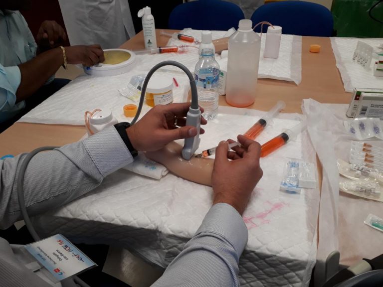
The EVF How Plus Sclerotherapy took place on the 21st and 22nd of March at the Hospital Central de la Cruz Roja in Madrid.  The EVF (European Venous Forum) is an international scientific society very involved in the training and dissemination of knowledge among phlebologists. Lourdes Reina tells us with words and images how these conferences were developed:

“46 ultrasound-guided sclerotherapy treatments were carried out. All complex cases, many of them varicose veins, pelvic varicose veins and advanced stages of chronic venous disease. Several cases of patients with healed or active ulcers were treated. Seven expert teachers and 12 students came from all over the world (South Africa, Turkey, Venezuela, Brazil, Serbia). All of them are physicians dedicated to Phlebology, with interest in expanding their knowledge in Sclerotherapy.   Theoretical lectures were given on technical aspects, complications, C1, varicose vein recurrence and of course an update on the treatment of venous ulcers. I gave a talk on venous ulcer treatment. I stressed the importance of carrying out a venous echoDoppler as soon as possible to look for the presence of superficial venous reflux and rule out the existence of extensive deep venous disease, in order to apply endoluminal therapy in the first two weeks of the ulcer ocurrence, with the aim of eliminating superficial venous reflux. I discussed the advantages of endoluminal therapy versus stripping surgery in patients with wounds and the advantages of sclerotherapy versus other endoluminal techniques. I also commented on the trend in the indication of endothermal techniques or sclerotherapy depending on the patient profile according to the experts.
 

Dear Lourdes, it is a great pleasure that our teams can work together to improve the lives of people with venous leg ulcers!


Definitive program EVF HOW Plus Course Sclerotherapy Madrid 21-22 March 2019

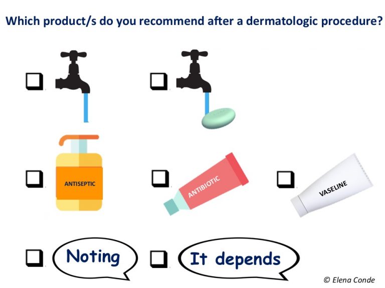
Why do we continue to use topical antibiotics in clean wounds after dermatologic procedures?

The unnecessary use of topical antibiotics in clean acute wounds is an issue I spoke about 4 years ago in the post How do we manage clean wounds secondary to dermatological procedures? I have been encouraged to write about it again after reading an interesting article recently published in the journal Dermatologic Surgery: “Variability in wound care recomendations following dermatologic procedures”.

The authors of this article perform a search on the recommendations after interventional procedures (biopsies, extirpations, curettage, cryotherapy, electrocoagulation, among others) that different dermatological centers have available on the Internet for patients. They analyse 169 protocols, mainly from US centres65% of them recommend the use of several topical products, which can cause doubts to the patient when choosing which one to use (and if there is an antibiotic in the list it is not difficult for the patient to think that this is the best option). Among the recommendations of the majority of protocols (84%) is the application of petrolatum-based products. However, it is striking that almost half of them (43%) propose the use of topical antibiotics, when the scientific evidence tells us that these drugs are not indicated in the management of these clean acute wounds. In addition to not representing a benefit to prevent infection or to accelerate healing in comparison with petrolatum- based products, their use can be related to bacterial resistance, development of allergic contact eczema and toxicity in fibroblasts and keratinocytes. In other words, not only do they not produce benefit, but they can also trigger complications that slow healing. In fact, certain topical antibiotics, such as neomycin, top the list of major allergens associated with allergic contact dermatitis.

Considering that there is no universally standardized protocol for the treatment of these post-surgical wounds, we should not be surprised by the heterogeneity of the recommendations found in this study. In fact, the same search carried out in Spanish would also show a great variability between centres and a widespread use of topical antibiotics.

But the problem of the absence of agreed and standardized treatment guidelines is not limited to wounds after dermatological procedures, but to clean acute wounds in general.  While there are multiple clinical guidelines and consensus documents for the treatment of chronic wounds, documents with evidence-based recommendations for the management of acute wounds are scarce. In this context, a multidisciplinary group of wound experts from the Netherlands reviewed the available evidence and published in 2015  a guideline on the management of acute wounds (Ubbink 2015). In the absence of scientific evidence on any point, the authors of the paper reflect their opinion as experts. I like the practical and concise approach of these guidelines, the recommendations of which coincide for the most part with the daily clinical practice of our practice. Among others, we find the following recommendations, to which I add in italics my personal opinion:

  1. Sutured wounds in aseptic conditions, as occurs in dermatological procedures such as biopsies and larger or smaller removals, do not require cleansing and the use of antiseptics, as the available evidence does not show that these measures reduce infection rates.
  2. If we leave a wound open for closure by secondary intention, recommendations will vary depending on the size and thickness of the wound. Superficial or small sized wounds would not need any coverage. As I commented in the post “Compressive therapy after dermatological surgery on the leg“, if no contraindication exists, as in any leg wound, therapeutic compression, adapted to the patient’s tolerance, helps to reduce inflammation to accelerate the healing of the lesions in this location.
  3. If the wound is closed and dry, covering it does not reduce the risk of infection and dressing changes can be painful. In addition, in sebaceous areas, such as the nose, occlusion or the use of petrolatum-based products can produce excess moisture, and consequent inflammation, with the possibility of delayed healing. In the case of mild exudate, the patient can use a conventional gauze dressing to absorb it and avoid friction with clothing. If the exudate is greater, a more absorbent dressing should be selected. No dressing has shown superiority over others. One type of wound that we let close by secondary intention is the graft donor site. As I commented in the post “What dressing should I choose to cover the graft donor site?” there are different options to adapt to the needs of the patient and the wound. The dressing that we use most in acute wounds is alginate, because of its hemostatic and  absorbent power and its capacity to form “pseudo-physiological” scabs (see post “Why do we use so many alginate fibre sheets in our wound clinic?”).
  4. Although it is a recommendation that we usually make in all cases, cleansing of acute wounds is also a controversial issue, since, as we saw in the post “The art of wound cleansing“, it does not seem to be associated with a lower rate of infection or other benefits in healing. In fact, the authors of this paper only recommend cleansing dirty wounds with warm tap water by gentle irrigation. Isn’t physiological scabbing the best coverage for a clean wound to heal?

With all this we have just commented on, it seems clear that we should not recommend mupirocin ointment to a patient after curettage and electrocoagulation of a skin lesion. However, is it better to recommend wound cleansing with soap and water and  vaseline every day or not to do any treatment? Is it better to apply vaseline or a repair cream with hyaluronic acid or zinc (see posts “Reasons for the hyaluronic acid boom in wound healing“, “Why do we use topical zinc in wounds and perilesional skin“)? What a dilemma… We need comparative studies that allow us to identify the best wound care protocol for clean acute wounds.

What is clear is that for these patients we must recommend sun protection during several months to reduce the risk of developing hyperpigmentation in the treated area.

In addition, since we know that a wound that does not close or causes pathological scarring (hypertrophy or keloid) is characterized by an abnormally prolonged inflammatory phase, the patient must avoid anything that may prolong that inflammatory phase. Therefore, other recommendations such as quitting smoking or avoiding activities that may produce tension at the edges of the wound will also facilitate healing.

After these dermatological procedures, the patient must receive clear instructions on what to do and what not to do, must be informed of possible complications that may appear, such as infection, and must ask any questions that arise. It is important for the patient to know that any healing process involves the presence of an inflammatory phase, the signs of which can sometimes be confused with those of infection. Well-informed patients have more control over their health and, consequently, their health will improve!

Gauzes soaked in astringent and antiseptic products for wounds and perilesional skin

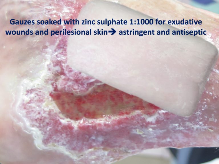
I’ve wanted to talk about this “dermatological” practice for a long time. I say “dermatological” because astringent and antiseptic products are a very widespread and effective practice in the treatment of exudative dermatoses. How useful is 1:1000 zinc sulphate solution in the vesicles and erosions of acute dermatitis or 2% sodium borate solution in diaper dermatitis! In the wound healing field there is very little described about the benefit of these solutions, but in our practice how we like zinc sulfate foments in exuding wounds and their macerated and eczematized perilesional skin!

As many of you will not be familiar with these treatments, we will start the explanation from the basics.


How can an astringent and antiseptic solution be applied?

A compress soaked in the solution is applied to the skin. The prolonged contact of this compress with the skin facilitates the penetration and, therefore, the action of the active principle of this solution. The adjective astringent refers to the drying action of this solution and antiseptic to its destructive or inhibiting power of different microorganisms, with low toxicity for our cells.

These aqueous solutions are obtained by dissolving the powdered presentation of the active ingredient in water, something that, depending on the substance, can be done by the patient himself or has to be prepared by the pharmacist.

A small reminder for those of you who are not clear about the indication of the different pharmaceutical forms: in very exudative lesions or in folds, powders in aqueous solutions must be applied to favour their drying, while in very dry lesions, ointments must be used to increase their absorption and hydrate the area. Creams are between these two extremes and their characteristics vary according to the percentage of water and fat they have.


What types of solutions with these characteristics can we use?

If one wants to review the astringent and antiseptic solutions, references in articles and books on wounds and healing are practically non-existent, so one must go to the dermatology or pharmacology manuals.

In fact, I have only found a couple of articles that comment on the interest of potassium permanganate in wounds.1 Of the rest, nothing, but here I present those that we use most frequently in exudative lesions in dermatology.

  • Zinc sulphate: this is the one we mainly use in our clinic, where we see predominantly leg wounds. Zinc can be combined with copper and aluminium to enhance its action. The antiseptic action of these three metals is due to their combination with the sulfhydryl group of certain enzymes, thus preventing the proliferation of microorganisms that depend on sulfur oxidation. These metal ions bind to human proteins, modifying their structure and causing them to precipitate. At low concentrations (1:1000), this denaturation and precipitation of proteins means that the ions do not penetrate deep tissues and their astringent action is produced. However, the high concentration of these metals is cytotoxic, as it damages the membranes and intracellular structures, and has a caustic activity.1
  • Boric acid: This acid has a predominantly antifungal action. It is not a fungicide, but slows fungal growth. In aqueous dilutions it increases its bacteriostatic power.1 There is a lack of evidence to support its use, but experience tells us that it is very interesting for very wet lesions where there is a risk of Candida We normally use 2% sodium borate, which we obtain by masterful formulation.
  • Potassium permanganate: as I mentioned, this is the only one of which there are punctual references in the scientific literature for its use in wounds. I have never used it. Its antiseptic action is due to its oxidizing power.1 By releasing oxygen, it oxidizes the nucleus and intracellular substances of the bacteria, and it also has astringent and deodorant power. To avoid irritation or even burns, it should be used at low concentrations, usually 1:10000.

In published cases, predominantly in cellulitis and acute eczema, which show the benefit of potassium permanganate foments, a steroid cream is also used, so it is difficult to determine the actual benefit of the solution.1

There is controversy regarding its interest in wounds, as many practitioners consider it to be an outdated practice and argue that there are other products that are safer and more effective.2


How are these solutions applied?

After soaking the gauzein the solution, we keep it on the wound and perilesional skin for approximately 10 minutes. Is this time sufficient? Wouldn’t longer contact be interesting? The problem with keeping the gauze longer is that it can lead to excessive absorption of the fluid by the tissue and worsen the exudate and maceration.

We use 1:1000 zinc sulfate in our clinical practice. To obtain this we dissolve a sachet of 1 g of zinc sulphate in 1 litre of water.

The ideal would be an application every 12 hours, as recommended by dermatologists for acute exudative lesions. However, in leg wounds, except in cases where the patient wears a compression stocking, the frequency of application must be adapted to that of the change of bandage. It is time to remember that if the wound exudate is not controlled, with secondary damage to the perilesional skin, the first thing to do is to think about what is failing: is the compression adequate? does the patient comply with the anti-edema measures we recommend? are there signs of infection? is the dressing adequate?

The combined application, first the astringent solution (in our case usually zinc sulfate) and then the topical corticosteroid cream, is something very used in our wound clinic, with excellent results. This is a widespread practice among dermatologists, especially in acute eczema, but isn’t the eczematized perilesional skin of our venous ulcer patients a dermatological problem that can benefit from this strategy?

As we commented in the post “Why do we use topical zinc on wounds and perilesional skin”, while zinc sulphate is used predominantly as a drying and antiseptic, zinc oxide is of greater interest in promoting healing. Therefore, after gauzes soaked with zinc sulphate, we apply zinc oxide barrier cream to the edges of the wound, which in addition to protecting the skin also promotes its repair. As you can find in this post about zinc, the combined application of topical corticosteroids and zinc oxide increases the beneficial effect of zinc.


Undoubtedly, the use of these solutions in wounds and perilesional skin is a therapeutic strategy with little evidence. Do you have any experience?


References:

  1. https://en.wikiversity.org/wiki/Antimicrobial_Agents_I
  2. Anderson I. Should potassium permanganate be used in wound care? Nurs Times. 2003 Aug 5-11;99(31):61. 
  3. Using potassium permanganate for wound cleansing. Readers’ Questions. Journal of Wound Care1994 3:4,

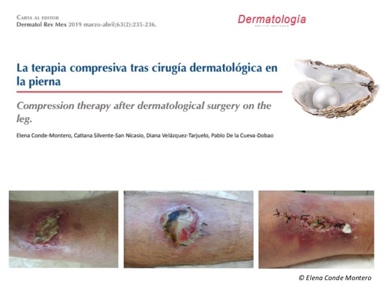
Compression therapy after dermatologic surgery on the leg

The truth is that I could also have titled this pearl “Compression therapy beyond venous leg ulcers”. In fact, as I have commented in different posts, if there is no contraindication, in any leg wound (post-surgical, post-traumatic, secondary to vasculitis…),medical compression, adapted to patient’s tolerance and needs, helps to reduce edema, inflammation and, consequently to accelerate healing (“Compression is key in to treating leg wounds”).

However, I have decided to talk specifically about compression therapy after dermatologic surgery because it is not a generalized practice among dermatologists and it should be, as you can read in the Letter to the Director that we have published in Revista Mexicana de Dermatología.

COMPRESSION IS THE BEST ANTI-GRAVITY AND, THEREFORE, ANTI-INFLAMMATORY TREATMENT OF LEG WOUNDS (not only venous ulcers) AS LONG AS NO CONTRAINDICATION EXISTS.

17 terapia compresiva

Therapeutic Pearl: Punch grafting in suboptimal wound beds

I have already talked about this topic in different blog entries (Punch grafting= a classic back in fashion, Interest of early punch grafting in Martorell hypertensive ischemic ulcer).

But I have selected it as a therapeutic pearl because we have just published an international consensus document (embedded at the end of the post and downloadable here) in which this advanced treatment for stagnant wounds is included, even if the wound bed does not present optimal conditions.

What I have already commented in previous occasions is perfectly reflected in this document:  In cases where the wound bed does not present with the optimal granulation tissue to receive skin grafts (for example, if stagnant slough is present), the ulcer may benefit from autologous punch grafting. Although some pinch or punch grafts may not adhere to the wound bed, they release growth factors, signal molecules and cells that enhance epithelial resurfacing and reduce pain”.

I take this opportunity to encourage you to read this multi-perspective and holistic paper on the diagnostic and therapeutic challenge of “super chronic” wounds that we often manage in our clinic – Remember that before treating a wound you must understand the patient and the factors that are preventing wound closure!

jowc.2019.28.sup3a.s1

Distraction as analgesic strategy during dressing changes

Professionals  involved in wound management know that dressing changes represent an enormously stressful factor for patients, mainly because of the associated pain. Pain has a sensitive component (the intensity of pain experienced by the patient), an affective component (emotional impact), and a cognitive component (thoughts about the experienced pain).1

It is not uncommon for painful experiences during wound manipulation to be recorded as traumatic memories. Fear of pain during treatment produces anticipatory anxiety in patients, which greatly limits their quality of life. In addition, this anxiety can condition the evolution of the wound, as it may cause patient avoidance of invasive interventions that promote healing, such as sharp debridement.

Commonly, when we think of analgesia, just systemic pharmacological treatment options come to mind. However, we are not always so aware of the adverse effects of these drugs, which is fundamental considering that patients with chronic wounds are usually older, with multiple comorbidities and multiple medications.

In previous entries I have talked about local analgesic treatments that we use daily in our clinical practice, such as EMLA cream, sevoflurane in irrigation or punch grafting. However, there are non-pharmacological and non-invasive techniques, some very easily applicable in any wound clinic, which can be very beneficial as an adjuvant treatment in our patients. Since it is a topic that I have just spoken about this weekend at the XIV meeting of the Dermatology and Psychiatry group of the AEDV, I am going to dedicate this post to it.

The benefit of these non-pharmacological analgesic strategies has been studied predominantly in burn units and children.

The techniques that we mostly use in wound clinics, even without being aware of it, are those of distraction.

Sleeping venus. Annibale Carracci. 1603. Museé Condé, Chantilly

Distraction aims to decrease pain by diverting attention away from the procedure being performed.1 This is explained by the pain gate theory: sensory stimuli from the new focus compete with painful stimuli for brain transmission, thus pain threshold increases. In addition to reducing pain and anxiety, distraction can reduce the time of interventions, as the patient collaborates more.

Distraction can also be divided into active and passive. Active distraction includes methods such as video games, virtual reality, active music therapy, controlled breathing, conversation, relaxation. Passive distraction techniques include listening to music or watching television.1,2

I have already dedicated a post to music (What if we heal with music?) But I want to talk about it again because the music in our clinical practice can be very profitable, we don’t have to limit ourselves to passive listening. The ideal situation would be to have a music therapist by our side. As this is normally impossible, we can encourage patient distraction by inviting them to choose the type of music they want to listen to, to sing their favourite songs or to take deep breaths accompanying the rhythm of the music. Music can also be an excuse to talk with them about musical tastes. It is important to remember the distracting power of conversation, which we use daily in our clinic.

The studies conducted, predominantly in children, point to a greater benefit of active methods. However, although conversation with parents is not as distracting as video games, it is effective in reducing anxiety and pain perception.2 If several professionals participate in dressing changes , it is very interesting that one engages in active distraction, predominantly with conversation.

Regarding innovative techniques, virtual reality is proving to be a very useful distraction technique, producing in the patient the illusion of being immersed in a completely different place.1,3 Virtual reality distraction, once developed and implanted in a wound centre, could be easily applicable in clinical practice, without much added effort or associated time.

Distraction by skin hyperstimulation, which has been predominantly described to relieve pain during injections,4 can also be very useful during invasive procedures in wound healing when there are several professionals or family members. There are different techniques, but we usually use pinching or pressure on the thigh or arm. The nerve impulses that we produce with this stimulation, if they are intense enough, can alleviate pain sensation.

If we review the published clinical trials on non-pharmacological strategies for pain control in patients with wounds, we find few papers, most of them on children and patients with burns, with small sample sizes and multiple limitations. In addition, many studies use combined techniques. All this makes it difficult to draw conclusions on the usefulness of each technique for the different types of wounds. Among the methods that seem to be most useful, in addition to distraction techniques, among which virtual reality stands out, is hypnosis.1

Hypnosis is a tool to alter the state of consciousness and to achieve a more suggestible situation.1 Basically, it is just another distraction technique.

In this state of unconsciousness, pain reduction would be achieved through cognitive changes that would alter affective states associated with pain (as we said at the beginning of the post, pain has a sensitive, cognitive and affective component). There are different schools and methods.5,6

Le Sommeil d Endymion. Anne-Louis Girodet. 1791. Museo del Louvre

One of the protocols most used by professionals who use hypnosis in patients with wounds is the so-called rapid induction of analgesia”(RIA),7 often with modifications, which achieves muscle relaxation and analgesia in a short period of time. Many professionals combine methods, such as relaxation, and use the suggestive force of reassuring words to generate positive sensations in the patient. It should be borne in mind that many people are not suggestible, so hypnosis in them is not effective.

Although the mechanism of pain reduction through hypnosis is not clear, associated neurophysiological changes have been detected, such as inhibition of nociceptive signals in the somatosensory cortex and modifications in the limbic system.5

To finish this entry on pain, a brief comment on its registration. In order to measure pain, different scales and questionnaires have been validated for clinical use and research. The visual analogue scale (VAS) is theoretically the best known but, in fact, it is the least used, as adaptations are normally used to facilitate its use, adding expressive faces or numbers. If we are purists, VAS for pain is a 10 cm line without marks or numbers. At one end there is “no pain” and at the other end there is “maximum pain”. Since patients with wounds are, in a high percentage, elderly people, this scale may not be understood. Consequently we use the numerical scale, visual or verbal, so that the patient expresses his perception of pain from 0 (no pain) to 10 (worst pain imaginable). When asked, we have to clarify what type of pain we want him to score: basal, irruptive (pain peaks) or procedural (pain associated with the procedure). In research studies it is important to evaluate anxiety with scales or specific questionnaires, since pain and anxiety are intimately linked in our patients with wounds.

Which techniques do you find most useful in your practice?


Referencias:

  1. Scheffler M, Koranyi S, Meissner W, Strauß B, Rosendahl J. Efficacy of non-pharmacological interventions for procedural pain relief in adults undergoing burn wound care: A systematic review and meta-analysis of randomized controlled trials. Burns. 2018 Nov;44(7):1709-1720. 
  2. Inan G, Inal S. The Impact of 3 Different Distraction Techniques on the Pain and Anxiety Levels of Children During Venipuncture: A Clinical Trial. Clin J Pain. 2019 Feb;35(2):140-147. 
  3. Scapin S, Echevarría-Guanilo ME, Boeira Fuculo Junior PR, Gonçalves N, Rocha PK, Coimbra R. Virtual Reality in the treatment of burn patients: A systematic review. Burns. 2018 Sep;44(6):1403-1416.
  4. Granados Navarrete I, Abril Sabater I, Alcaraz Busqueta F, Mañé Buixo N, Padilla Ruiz J, Real Gatius J, Solano Pallarés M, Yuste Giménez, E. Una actuación de enfermería: intentar aliviar el dolor en las punciones de hemodiálisis. Revista de la Sociedad Española de Enfermería Nefrológica. 2005; 8(3): 55-60.
  5. Jafarizadeh H, Lotfi M, Ajoudani F, Kiani A, Alinejad V. Hypnosis for reduction of background pain and pain anxiety in men with burns: A blinded,randomised, placebo-controlled study. Burns. 2018 Feb;44(1):108-117.
  6. Provençal SC, Bond S, Rizkallah E, El-Baalbaki G. Hypnosis for burn wound care pain and anxiety: A systematic review and meta-analysis. Burns. 2018;44(8):1870-1881. 
  7. Barber J. Rapid induction analgesia: a clinical report. Am J Clin Hypn. 1977 Jan;19(3):138-45.