Home Blog Page 6

Therapeutic pearl in post-surgical wounds

If anything makes me adore pearls is the elegance with which they help you succeed in any situation. In fact, the success  in our clinic is based on these therapeutic jewels.

So I have decided to start making a necklace of therapeutic pearls, which will be greatly enriched by your contributions.

And is there a better way to start this section of the blog than with an official pearl? I say official because we have just published Punch grafting in post-surgical wounds (link)(doi: 10.1016/j.jaad.2019.02.060.) in the Therapeutic Pearls section of the Journal of the American Academy of Dermatology 

Here I share the article, but if you want more details about this pearl, I invite you to read the post “Punch grafting in post-surgical wounds

“Punch grafting in post-surgical wounds” ©2019. This manuscript version is made available under the CC-BY-NC-ND 4.0 license http://creativecommons.org/licenses/by-nc-nd/4.0/


Punch grafting: a classic back in fashion

The 8th Congress of the SEHER (Spanish Wound Healing Society) has just been held in Madrid https://www.congresoheridas.com/index.php/programa. Once again, this scientific meeting has been a meeting point for colleagues and friends. The star novelties of last year’s congress, such as topical sevoflurane in wounds or the App Wound-Registry have been kept among the leading topics in this edition.

It was a pleasure to moderate and participate in a session dedicated to Dermatology in the world of wounds. This year I decided to talk about the interest of early punch grafting in arteriolopathic ulcers, something I love and what I have recently talked about in the blog (“Large leg wounds after mild trauma”)

But what I really loved was to hear and see the excellent results of other colleagues with punch grafting in recalcitrant wounds. This simple, economic and very effective treatment has more and more presence in posters and communications of this congress.

To encourage you to start applying the technique and not to be discouraged when the first dressing changes do not look the way you expected, here I answer the 10 questions that arise most frequently.


  1. Are these grafts similar to epidermal grafts obtained with the marketed product?

No, they are different. Punch grafts consist of epidermis and superficial dermis, therefore, in the donor site there is a  point-like bleeding and when placed on a wound bed in good conditions is produced, graft taking is expected to occur. Epidermal grafts only consist of epidermis, so there is no bleeding after obtaining them, but when they are placed in the wound there is no graft taking and epithelialization is promoted by cells and growth factors released in the bed. I encourage you to read the blog entry in which I explain it in detail: “Differences between epidermal and dermo-epidermal punch grafts”


  1. Should I suspend oral anticoagulants before performing the technique?

Since this is a surgery with low bleeding risk, we usually do not suspend them. However, before performing the grafts it is important to ensure that there is good control of the anticoagulation state (INR must be in the expected range).


  1. How do I obtain grafts from the donor site?

We usually obtain them from the antero-lateral aspect of the thigh. We infiltrate tumescent anaesthesia with 1% lidocaine diluted with physiological serum.

The grafts may be obtained with punch, curette or scalpel blade. This last system is the one we currently use most frequently.

It is a technique very well tolerated by the patient and we carry it out in the consulting room.

See post “Types of skin grafts to cover chronic wounds: Which one should you choose”


  1. What is the expected evolution of the donor site?

Let´s see an example. Immediately after obtaining the grafts, we cover the donor site with calcium alginate and use gauze as a secondary dressing. In this case the first dressing change was carried out on the third day and the donor site as covered again with alginate. One week after the procedure, with scab formation, it was recommended the daily application of a repairing cream with zinc (See post “Why do we use topical zinc on wounds and perilesional skin?”)

In the entry “Which dressing should I choose to cover the skin graft donor site?” you can find more information about the local care of the donor area.


  1. What dressing should I use to cover the grafted wound?

We usually cover the grafted area with sheets of calcium alginate, as I have told you in the entry “Why do we use so many alginate sheets in our wound clinic?”. Another option is using interfaces (we mainly use a mesh with lipido-colloid technology and anti-metalloproteinases activity) as primary dressing, and alginate sheets as secondary dressing. In special locations, such as the scalp, flexible foam dressings with adaptable edges are useful to avoid bulky bandages.


  1. When is it useful to combine punch grafts with negative pressure therapy?

Negative pressure therapy can help improve the wound bed before graft coverage, but it is also very interesting for promoting graft attachment (especially if wound bed conditions are bad or in special locations, such as the Achilles tendon). You can read more at “Punch grafting and Negative Pressure Therapy: A Successful Couple” 

  1. What types of wounds can benefit from this technique?

Any wound in which epithelialization is not achieved, provided that there are no signs of infection, the etiological treatment has been optimized and the patient is not a candidate for other reconstructive surgical procedures.

The spectrum of arteriolopathic ulcers, which includes Martorell ulcer and post-traumatic wounds in the context of age-associated arteriolosclerosis, this early treatment is of high interest for, in addition to promoting healing, reducing pain and avoiding lesion progression (See entry: “Large leg wounds after mild trauma”)


  1. Is it possible to cover sloughy wound beds with punch grafts?

The ideal situation is to graft optimal beds with red granulation tissue. However, our patients are usually older, with multiple comorbidities, associated pain and anticipatory anxiety for dressing changes. Although we try to improve wound bed tissue as much as possible, it is not always easy to achieve optimal conditions for graft coverage. However, experience tells us that, despite devitalized tissue in the bed and the absence of complete graft taking, the wound benefits from this coverage. Punch grafts behave like independent micro-islands, so those that do not take do not affect the rest and are also beneficial because they can release cells and growth factors interesting for healing. In addition, the analgesic action of these grafts must be underlined, which, as we observed in our clinical practice, is independent of the percentage of graft taking. See post: “Interest of early punch grafting in Martorell hypertensive ischemic ulcer”.


  1. How is the expected evolution of the grafted wound?

It varies greatly depending on the characteristics of the wound bed, the wound evolution time, the co-morbidities of the patient and the immobilization of the grafts the first few days after coverage. This immobilization is achieved with local pressure. In leg wounds we put compression bandages and recommend rest with the limb elevated during one week, which is essential during the first 3-4 days.

It is also key to space dressing changes as far as possible and to be as conservative as possible in wound bed cleansing, without removing the grafts that have not taken or the gelled or crusty material that appears between them. IN EACH DRESSING CHANGE WE REMOVE WHAT IS NOT INTERESTING FOR THE WOUND, BUT ALSO GROWTH FACTORS AND CELLS THAT ARE PROMOTING HEALING!

This is the clinical aspect during weekly dressing changes after punch grafting a venous ulcer in the inner aspect of the right leg, of three years of evolution. The whitish colour in the perilesional skin is due to the zinc barrier product we use (See post “Why do we use topical zinc on wounds and perilesional skin?”)


  1. If no graft taking has occurred , is it a total failure?

Quite the opposite. In fact, on many occasions we graft wounds with the sole aim of reducing pain. In addition, when we graft beds with abundant slough, even though no graft taking exists, neoangiogenesis is produced, which improves granulation tissue, and cells and growth factors that promote wound contraction are released. In these cases, we have to perform several sessions of punch grafting to obtain complete epithelialization, something very well accepted by the patient.

I would like to end with heartfelt congratulations to my great colleague and friend, Laura Pérez Jerónimo, for the second prize in Clinical Cases that she received at the 8th SEHER Congress. As could not be otherwise in our practice, this case was treated with punch grafts. You can see her in the photo with Alicia Pérez Vázquez and Lorena Recarte Marín, excellent colleagues and friends too. Congratulations, team!

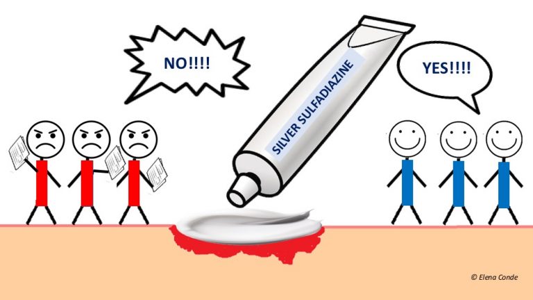
Silver sulfadiazine: adored by some, detested by others.

Silver sulfadiazine cream (better known by its commercial names) remains, since the 70s, in the top of products used in wound healing, predominantly in superficial burns. It seems that the opinion of professionals on this product is divided into two camps: supporters and detractors. Although it is a product that I do not use in my clinical practice, its presence in various presentations during the Journées Cicatrisations 2019 has encouraged to dedicate a post to it:)


What is silver sulfadiazine?

Silver sulfadiazine is a combination of silver nitrate and sulfadiazine, so its bacterial effects are due to the action of these two active principles.

We have already dedicated a post to silver (“Silver in skin wounds”). Sulfadiazine is a type of sulfamide, known traditional broad-spectrum antibacterial (Gram +, Gram – including Pseudomonas, anaerobes). This chemical has a bacteriostatic action, i.e. it prevents the reproduction of bacteria by interfering with their folic acid synthesis. Therefore, when combined in a cream, the bacteriostatic power of sulfadiazine and the silver bactericide effect act in synergy. In short, it is a mixture of a topical antibiotic with silver. Its usual presentation is cream, at a concentration of 1%. The general recommendation is to apply it daily after cleansing the wound to avoid the formation of a pseudo-scab.

I imagine that many of you will already have the following question in your head…


But is it advisable to apply topical antibiotics on wounds?

Although they are commonly used in acute and chronic wounds, the available evidence does not allow us to recommend them. In fact, in clean acute wounds, such as those secondary to dermatological procedures, although the use of topical antibiotics is not indicated, it is a very widespread practice (see post: “How do we manage clean wounds produced after dermatological procedures?“). In chronic wounds, in addition to not having a clear effect on bacteria organized and protected in biofilms, they can produce bacterial resistance and trigger allergic reactions. Returning to the specific topic of silver sulfadiazine, harmful effects on healing have been shown in vitro and in vivo, such as altered activation of macrophages and cytotoxicity on keratinocytes and fibroblasts.1 Allergic contact dermatitis from silver sulfadiazine, either by allergic reaction to silver or sulfadiazine, is not uncommon.2 If a wound worsens after starting treatment with silver sulfadiazine, with increased pain, exudate, and perilesional redness, it may be clinically suspected. This adverse effect is confirmed by epicutaneous tests (patch test). Nitrofurazone is another topical antibiotic widely used in burns and other wounds, which dermatologists know well because it is also responsible for many hypersensitivity reactions.

In order to avoid the possible effects of systemic absorption of silver sulfadiazine, it is necessary to avoid its application in very extensive wounds during prolonged periods of time, especially in patients with renal or hepatic insufficiency. In addition to the potential accumulation of silver in different organs, sulfadiazine could produce blood alterations such as methemoglobinemia, haemolysis, leukopenia or hyperbilirubinemia. With the use of silver sulfadiazine severe cutaneous drug reactions have also been described, such as Stevens-Johnson syndrome.3 However, these adverse effects are considered rare, so silver sulfadiazine is considered a safe drug.


What do the studies conclude?

Most studies are conducted on splitl-thickness burns. However, before we focus on this group of patients, let´s see what has been published on silver sulfadiazine in venous ulcers, as there are many professionals who use it with this indication.

Two prospective series, with 64 and 70 outpatients each (in German and French respectively),3  analyse the number of patients with complete healing at 6 weeks of weekly application of silver sulfadiazine cream. These series found complete healing in 52 and 40 patients respectively, with loss of follow up of 7 patients in this second group. With regard to signs of infection, in the first study they were not detected. In the second study, while 29 patients presented infection data on the day treatment began, only 2 had these signs at 6 weeks. However, a randomized clinical trial with 86 patients with venous ulcers in which silver sulfadiazine is compared to placebo does not show significant differences in complete healing in the two groups.3

Now let’s focus on superficial burns, undoubtedly its most widespread indication. First of all, it must be stressed that the available evidence is scarce and of low quality. In animal models, with rats and pigs, experimental studies have been carried out to compare the benefit of silver sulfadiazine versus placebo (including paraffin gauze, gauze with saline serum, oily excipient without active principle) for the outcome variables “infection” or “complete healing”, with variable results.3

In humans, on the contrary, clinical trials essentially compare silver sulfadiazine cream with other products, as they consider the former as the standard of treatment and normally use it in the control group. Let’s see what these studies conclude in superficial burns in relatively small areas.

  • Non-occlusive petrolatum gel vs. silver sulfadiazine covered with gauze: a randomized clinical trial compares these two strategies, with daily cures, in two groups of 19 patients each. No significant differences were found in time to complete healing, infection or dermatitis signs.4
  • Silver sulfadiazine vs. paraffin gauze: one study included 15 superficial burns, dividing each wound into an upper half treated with paraffin gauze and the lower half with silver sulfadiazine. Paraffin gauze was changed every 5 days, while silver sulfadiazine was applied daily and also covered with paraffin gauze. Healing of the area treated only with paraffin gauze was faster.5
  • Silver sulfadiazine vs. hydrocolloid dressing: a randomized clinical trial included 50 patients. Burns in the hydrocolloid dressing group required fewer dressing changes and showed faster complete epithelialization with better appearance and pigmentation of the scar area. Patients reported less limitation in their usual activities and greater adherence to hydrocolloid treatment, benefits that add up to lower treatment costs.6
  • Silver sulfadiazine vs. honey: there is growing interest in the use of honey in healing, due to its potential debridement, anti-inflammatory and antibacterial activity. A recent systematic review7 includes 9 clinical trials comparing the use of honey and silver sulfadiazine in superficial burns. This study finds faster complete healing and a higher percentage of infection resolution in honey-treated wounds. However, it should be noted that these are small studies (between 27 and 110 patients included) very heterogeneous, with different age groups, variable outcome measures and duration of follow up.7
  • Silver sulfadiazine vs. polyhexanide/betaine gel: polyhexanide/betaine gel has a great interest in wound healing due to its antibacterial and moisturizing action. A randomized clinical trial involving 46 patients found no significant differences in time to complete healing, infection and costs between the group treated daily with silver sulfadiazine and that of polyhexanide/betaine.8


What do the position documents and clinical guidelines say?

As we have just seen, although the available studies are few, small in size, and heterogeneous in design, most show the absence of differences or even superiority of other treatments over silver sulfadiazine for superficial burns, in terms of infection and complete healing.9 However, if we do a quick search for position documents and clinical practice guidelines, we will find the widespread recommendation for the use of silver sulfadiazine cream in split- thickness burns. In the guideline published by the Japanese Burn Society in 2016,10 as well as in other clinical guidelines, it is underlined that its recommendation for use is made despite the absence of a good level of evidence.

What is your experience with silver sulfadiazine cream?


References:

  1. Punjataewakupt A, Napavichayanun S, Aramwit P. The downside of antimicrobial agents for wound healing. Eur J Clin Microbiol Infect Dis. 2019 Jan;38(1):39-54. 
  2. García AA, Rodríguez Martín AM, Serra Baldrich E, Manubens Mercade E, Puig Sanz L. Allergic contact dermatitis to silver in a patient treated with silver sulphadiazine after a burn. J Eur Acad Dermatol Venereol. 2016 Feb;30(2):365-6
  3. Miller AC, Rashid RM, Falzon L, Elamin EM, Zehtabchi S. Silver sulfadiazine for the treatment of partial-thickness burns and venous stasis ulcers. J Am Acad Dermatol. 2012 May;66(5):e159-65. 
  4. Genuino GA, Baluyut-Angeles KV, Espiritu AP, Lapitan MC, Buckley BS. Topical petrolatum gel alone versus topical silver sulfadiazine with standard gauze dressings for the treatment of superficial partial thickness burns in adults: a randomized controlled trial. Burns. 2014 Nov;40(7):1267-73. 
  5. Stern HS. Silver sulphadiazine and the healing of partial thickness burns: a prospective clinical trial. Br J Plast Surg. 1989 Sep;42(5):581-5.
  6. Wyatt D, McGowan DN, Najarian MP. Comparison of a hydrocolloid dressing and silver sulfadiazine cream in the outpatient management of second-degree burns. J Trauma 1990;30: 857-865.
  7. Aziz Z, Abdul Rasool Hassan B. The effects of honey compared to silversulfadiazine for the treatment of burns: A systematic review of randomized controlled trials. Burns. 2017 Feb;43(1):50-57.
  8. Wattanaploy S, Chinaroonchai K, Namviriyachote N, Muangman P. Randomized Controlled Trial of Polyhexanide/Betaine Gel Versus Silver Sulfadiazine for Partial-Thickness Burn Treatment. Int J Low Extrem Wounds. 2017 Mar;16(1):45-50.
  9. Chung JY, Herbert ME. Myth: silver sulfadiazine is the best treatment for minor burns. West J Med. 2001;175(3):205-6. 
  10. Yoshino Y, Ohtsuka M, Kawaguchi M, Sakai K, Hashimoto A, Hayashi M, Madokoro N, Asano Y, Abe M, Ishii T, Isei T, Ito T, Inoue Y, Imafuku S, Irisawa R, Ohtsuka M, Ogawa F, Kadono T, Kawakami T, Kukino R, Kono T, Kodera M, Takahara M, Tanioka M, Nakanishi T, Nakamura Y, Hasegawa M, Fujimoto M, Fujiwara H, Maekawa T, Matsuo K, Yamasaki O, Le Pavoux A, Tachibana T, Ihn H; Wound/Burn Guidelines Committee. The wound/burn guidelines – 6: Guidelines for the management of burns. J Dermatol. 2016 Sep;43(9):989-1010. 


How do we manage clean wounds secondary to dermatological procedures?

Dermatology is a specialty in which we perform daily invasive processes (cryotherapy, laser, electrocoagulation, biopsies, extirpations), with the consequent production of wounds that are considered clean lesions. If we analyse our daily clinical practice, type of dressing changes until complete re-epithelialization is very variable. A wide variety of recommendations may be found, such as cleansing with soap and water, chlorhexidine or povidone iodine, antibiotic ointment, vaseline products, occlusive dressings…

The use of antiseptics or antibiotics to prevent wound infection seems to give us peace of mind (Nijhawan, 2013). Many professionals recommend the use of topical antibiotics since their presentation in ointment favours healing in a moist environment. However, the use of these products in a prophylactic way is not indicated after dermatological clean invasive procedures (Levender, 2012).

The incidence of complications in dermatological surgery is very low, varying in the studies from 0.07 to 4.25% (Rogers, 2010; Lee, 2015). The use of antiseptics in dressing changes after these clean procedures does not represent a benefit and may be harmful in the healing process due to their cytotoxicity (Cho, 1998). On the other hand, the indiscriminate use of topical antibiotics, in addition to not having shown a benefit in reducing infection rates, contributes to the development of antibiotic resistance and the onset of contact dermatitis (Gehrig, 2008), with an increase in healthcare costs.

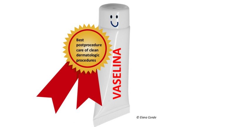
Topical vaseline products would represent a safe treatment option as they promote healing in a moist environment without the aforementioned inconveniences.

Different clinical trials and a systematic review have been carried out with the aim of determining the efficacy of antibiotic ointments, in comparison with vaseline, in the prevention of infection of clean surgical wounds (Saco, 2014; Lee,2015). Among the results and conclusions of these studies, the recommendation of the use of vaseline, and not antibiotic ointments, stands out for  healing of clean wounds after dermatological procedures. The application of vaseline would not be necessary in cases where occlusive dressings are used, since no statistically significant differences have been found on the risk of infection and the final result between adding or not adding ointments (vaseline alone or with antibiotic) under them (Dixon, 2006). However, the use of occlusive dressings implies a higher expense than the use of vaseline ointment and an adhesive dressing with gauze, and no significant benefit has been demonstrated from their use in this type of wounds (Ubbink, 2008).


What do you recommend? What is your experience?


References:

Cho CY1, Lo JS. Dressing the part. Dermatol Clin. 1998 Jan;16(1):25-47.

Dixon AJ, Dixon MP, Dixon JB. Randomized clinical trial of the effect of applying ointment to surgical wounds before occlusive dressing. Br J Surg 2006;93:937-943.

Gehrig KA, Warshaw EM. Allergic contact dermatitis to topical antibiotics: Epidemiology, responsible allergens, and management. J Am AcadDermatol 2008;58:1-21.

Lee DH, Kim DY, Yoon SY, Park HS, Yoon HS, Cho S. Retrospective Clinical Trial of Fusidic Acid versus Petrolatum in the Postprocedure Care of Clean Dermatologic Procedures. Ann Dermatol. 2015 Feb;27(1):15-20.

Levender MM, Davis SA, Kwatra SG, Williford PM, Feldman SR. Use of topical antibiotics as prophylaxis in clean dermatologic procedures. J Am Acad Dermatol. 2012 Mar; 66(3):445-51. Epub 2011 Aug 6.

Nijhawan RI, Smith LA, Mariwalla K. Mohs surgeons’ use of topical emollients in postoperative wound care. Dermatol Surg. 2013 Aug;39(8):1260-3.

Rogers HD, Desciak EB, Marcus RP, Wang S, MacKay- Wiggan J, Eliezri YD. Prospective study of wound infections in Mohs micrographic surgery using clean surgical technique in the absence of prophylactic antibiotics. J Am Acad Dermatol 2010;63:842-851

Saco M, Howe N, Nathoo R, Cherpelis B. Topical antibiotic prophylaxis for prevention of surgical wound infections from dermatologic procedures: a systematic review and meta-analysis. J Dermatolog Treat. 2014 Apr 8.

Ubbink DT, Vermeulen H, Goossens A, Kelner RB, Schreuder SM, Lubbers MJ. Occlusive vs gauze dressings for local wound care in surgical patients: a randomized clinical trial. Arch Surg. 2008 Oct;143(10):950-5


Myofibroblast: the great ignored in wound healing

And why did I just remember the myofibroblast? Because a lot has been said about it in the Journées Cicatrisations 2019 (National Congress of the French Society of Wounds and Healing) in Paris. Once again this year, it has stood out for the high quality of the programme and the excellence of the speakers.

As it is typical in this scientific meeting, it has been discussed about both innovation and tradition. In this line, punch grafting, a technique that was described more than 150 years ago, has been again the subject of different sessions. It seems to be widely accepted that this procedure stands out, apart from its acceleration of epithelialisation, its simplicity, its analgesic power (very interesting in the Martorell ulcer) and its possibility of being combined with other techniques, such as negative pressure therapy. We cannot forget that the epithelialisation of a wound is produced by the linear proliferation of keratinocytes. One of the main obstacles is the senescence of these cells in long-standing wounds. That’s why we graft these stagnant wounds, voilà! I was delighted to see experiences similar to those we have in our practice ?.

Let’s go back to myofibroblast. To understand the behaviour of this cell, its name gives us many clues (myo=muscle, fibro=fibre). This is a cell that promotes the contraction and production of collagen fibres in the wound.

This contractile power is due to the fact that it is a type of activated fibroblast that expresses in its cytoskeleton α-actin, a protein typical of smooth muscle, fundamental in the contraction process of muscle cells.

Myofibroblast is one of the main stars in wound healing, since its deficit will obviously slow down wound closure. In elderly people, or people with comorbidities that prolong the inflammatory phase of healing, proliferation and activity of myofibroblasts may decrease, as they depend on cytokines and growth factors released by cells such as macrophages M2, whose number is decreased in chronic wounds.1,2

A short reminder about macrophages:

A macrophage is a type of white cell (monocyte) that has specialised in the ingestion and destruction of bacteria and cell debris (phagocytosis) in tissues. Depending on the signals they receive from the environment, they can be differentiated into type 1 macrophages (M1) and type 2 macrophages (M2). The activity of the former is essential for the organism protection against pathogens (as occurs in acute wounds) but, if maintained over time, the pro-inflammatory cytokines they release stimulate a proteolytic environment that prevents healing. M2 cells release anti-inflammatory molecules to promote tissue repair.


There’s no doubt that wounds need myofibroblasts. But watch out, not too many, not too long! Let’s see why I say this.

Below I have simplified what happens at the cellular level in an acute wound, which we academically divide into overlapping phases (hemostasis, inflammation, proliferation and remodeling). See post:”Brief overview of wound healing”. Complete epithelialization represents both the end of the wound and the birth of what is known as a scar. The healing process includes everything that happens from the appearance of the wound to the complete maturation of the scar.

Fibroblasts proliferate and become active myofibroblasts when inflammation decreases and the molecular microenvironment favours the production of granulation tissue. Myofibroblasts proliferation is then maximal to produce collagen and favour the contraction of the lesion.

As with the rest of the cells involved in the formation of granulation and epithelial tissue, as days go by and scar remodelling progresses, the programmed death (apoptosis) of myofibroblasts causes reduces their number in a few weeks.1

What can happen if the balance between cells, growth factors and extracellular matrix is altered and the activity of the myofibroblasts increases and is prolonged over time, months or years? An excessive contraction and fibrosis, that is to say, a pathological scar.

The remodelling phase of a scar is the least known, but it is essential for the extracellular matrix to reorganize, for type III collagen to become type I and for the tissue to gain the maximum elasticity possible. It has been shown that the density of myofibroblasts maintained over time produces excessive scarring, with its cosmetic and functional repercussion.1

Theory is fine, but how can we help in order to activate them in stagnant chronic wounds and stop them when we want to prevent or treat hypertrophic scars?


How to activate myofibroblasts in chronic wounds?

All strategies that reduce inflammation (control of comorbidities, appropriate etiological treatment, local treatment of the wound bed) will favour their proliferation and activation.  The mechanical stretching force produced by negative pressure therapy on the wound bed and edges is a good way to stimulate myofibroblasts.1


How to reduce its activity to prevent or treat hypertrophic and keloid scars?

There is growing interest in research into pro-myofibroblast factors, including the Transforming Growth Factor β1 (TGF β1), whose inhibition would prevent fibrosis.1 The problem is that this and other proteins are also involved in the proliferation, among others, of keratinocytes and endothelial cells, thus affecting general wound healing. Specific targets must therefore be sought.

Currently, silicone dressings and corticosteroid infiltrations are the measures most recommended in the guidelines and most used in clinical practice. Multiple mechanisms of action of silicone have been proposed, mainly silicone sheets, among them that moisture and occlusion it provides would simulate the protective characteristics of healthy skin and reduce the activity of myofibroblasts and collagen formation. In addition, consequent decreased tension from the sides of the scar can also slow down the activity of myofibroblasts, as these cells respond to mechanical forces. With respect to the corticosteroids, they have been shown to inhibit TGF-β1 expression, thus inhibiting the proliferation of myofibroblasts and favouring their apoptosis.

Other treatments, including cryotherapy, 5-fluorouracil (5FU) or bleomycin injections, laser, can also be very useful.3

However, rather than focusing on single treatments, the general recommendation is to use combined strategies, focusing on the importance of regenerative rehabilitation. What do I mean by this? That we have to optimize scar functionality, with physical treatments that favour its elasticity and reduce adhesions.

In another post we have already talked about the benefits of massage (“Compression therapy and scar massage in post-traumatic and post-surgical wounds“) but I would like to recall the interest of this and other physical therapies to promote the appropriate scar remodelling.

Any mechanical forces exerted on the skin, although minimal, affect the cells, which detect them and adapt to the change by modifying their activity. The healing process is strongly influenced by the effects of mechanotransduction (i.e., transformation of the biomechanical force on the cell membrane into intracellular molecules that act as second messengers). The effect of this mechanical force may be clinically observed in the characteristics of the scar. While it has been shown that stretching forces on a scar maintained over time increase TGF-β1 levels, activity of myofibroblasts and collagen formation, other forces, such as those produced during massage, can produce the opposite effect. However, optimal intensity, duration and frequency of this type of treatment are unclear.

Another interesting strategy is pressure therapy on scars, which has been studied predominantly in burns (wounds with well-known high risk of hypertrophy). The most widespread recommendation is to use devices or garments that exert a local pressure maintained between 20-30 mmHg, which is sufficient to overcome capillary pressure without the possible adverse effects of higher pressures. By decreasing local capillary flow, activity of myofibroblasts and collagen production is reduced.3

Returning to the interest of combined treatments, Peter Moortgat‘s team (whom I had the pleasure of hearing at the Journées Cicatrisations) has just published a randomised study to analyse the benefit of pressure therapy added to silicone occlusion.4 The clinical trial includes 60 patients with hypertrophic scars after burns of variable evolution time and compares the benefit of using a cohesive silicone bandage and silicone sheet after 6 months of treatment. No differences were found in colouring and subjective assessment by the patient, but in the compression bandage group greater scar elasticity and a better subjective score by the practitioner were found. The authors associate improvement with elevated and continued pressure on the scar area. Among the limitations of the study are the different characteristics between the groups, the different scar locations, and the short duration of the study (since alterations in scar remodelling may appear more than a year later).

More studies would be interested to answer multiple questions that arise: When should we start pressure therapy and how long should it be maintained? Which device is the most suitable for our patient?


References:

  1. Hinz B. The role of myofibroblasts in wound healing. Current Research in Translational Medicine 64 (2016) 171–177. 
  2. Shook BA, Wasko RR, Rivera-Gonzalez GC, Salazar-Gatzimas E, López-Giráldez F, Dash BC, Muñoz-Rojas AR, Aultman KD, Zwick RK, Lei V, Arbiser JL, Miller-Jensen K, Clark DA, Hsia HC, Horsley V. Myofibroblast proliferation and heterogeneity are supported by macrophages during skin repair. Science. 2018 Nov 23;362(6417).   
  3. Kafka M, Collins V, Kamolz LP, Rappl T, Branski LK, Wurzer P. Evidence of invasive and noninvasive treatment modalities for hypertrophic scars: A systematic review. Wound Repair Regen. 2017 Jan;25(1):139-144. 
  4. Moortgat P, Meirte J, Maertens K, Lafaire C, De Cuyper L, Anthonissen M. Can a cohesive silicone bandage outperform an adhesive silicone gel sheet in the treatment of scars? A randomised comparative trial. Plast Reconstr Surg. 2018. 

Compression is key to treating leg wounds

Some of you may have been struck by this title, which, instead of “venous ulcers“, includes the term “leg wounds”. Leg wound is a general and nonspecific concept, so it fits perfectly into the header of this post. Regardless of its cause, when no contraindication exists, a leg wound will benefit from compressive therapy.

I hope that reading this post will gain you confidence in compression therapy and become your main ally in the treatment of leg ulcers.


Why do leg wounds benefit from compression therapy?

manzana gravedad

The inflammatory process inherent in any leg wound involves an alteration in the microcirculation, with greater capillary filtration, and, due to the effect of the force of gravity, an increase in intravenous pressure. Therefore, despite the absence of reflux or an obstructive picture, a state of venous hypertension may develop, making healing difficult. The term “hydrostatic ulcers” has been proposed to refer to leg wounds of different aetiologies that meet this condition. That is, they do not meet the definition of venous ulcer but behave as if they were (and many seem to be clinically). This group includes wounds in patients with obesity, immobilization, traumatic wounds, secondary to vasculitis or occlusive vasculopathy… It is not uncommon for a patient to present several of these predisposing conditions together. However, the complexity that we find in our clinical practice should not modify the essence of our therapeutic attitude: in order to fight against venous hypertension, present to a greater or lesser extent in any leg wound, the promotion of venous return through compressive therapy is fundamental.

The effects of compressive therapy on leg wounds are, among others:

  • Decreased capillary filtration
  • Increased local lymphatic drainage
  • Reduction of inflammation
  • Increased arterial flow

These benefits justify the recommendation to use compression therapy, provided it is not contraindicated, in any leg with a wound. Here are some examples:


What about arterial component wounds?

As we have just pointed out, our patients usually present with comorbidities and the wounds usually have several associated components, such as arteriopathy.

As a principle, pressure exerted externally continuously must not exceed the intra-arterial and arteriolar pressure. This is why it has been established that compressive therapy is contraindicated if the ankle-brachial index (ABI)  is less than 0.5. However, this does not mean that patients with peripheral artery disease cannot benefit from compressive therapy. In fact, increased arterial flow has been shown in these patients with the use of stiff bandages or pneumatic compression devices. Stiff bandages and pneumatic compression, unlike bandages with elastic properties, only exert intermittent, unsustained pressure (during exercise and at scheduled intervals, respectively). See post Band and bandage: not the same thing

However, we must not forget that the ABI is an indicator of arteriopathy, not ischemia (real contraindication for compression). In addition, this rate may be falsely elevated by calcification of the arterial media. Therefore, as I have already commented in other entries, in cases of doubt (arterial calcification, diabetic patient), arterial risk must be defined by a combination of clinical examination (characteristics of pain and trophic alterations) and absolute measures of chronic ischemia (systolic pressure in the first toe and in the ankle).

With regard to the other absolute and relative contraindications of compression therapy, the recommendations vary depending on the clinical guideline. This lack of consensus and absence of specificity is summarized in a poster that was presented at the 5th WUWHS congress held in September in Florence and can be downloaded in http://www.icc-compressionclub.com/. In practice,  regarding diabetic patients with arteriopathy, neuropathy, heart failure, skin fragility and many other added risk factors, I believe that the key to success is a close follow-up, examining the limb in detail at each dressing change and, most importantly, listening to the patient.


Conclusions

  • Compression therapy is the anti-gravity treatment of leg wounds.
  • Provided that there are no contraindications for its use, a leg ulcer will benefit from the antihypertensive and anti-inflammatory effect of compressive therapy.


I recommend this monographic issue of compression therapy published in 2010 in the Journal of Wound Technology, which you can also access from the very interesting website of the International Compression Club. Had you ever heard about it?


Chronic venous insufficiency from a dermatological perspective

“Chronic venous insufficiency from a dermatological perspective. This was the title of my lecture at the 7th Congress of the Spanish Society of Wounds (SEHER) in Madrid. I had the pleasure of talking about the cutaneous impact of venous hypertension, beyond the venous ulcer.

Looking forward to the 8th edition of the SEHER Congress in 2019,  I am going to summarize the contents of my lecture in a short question-answer format:

– How does the skin respond to venous hypertension?

With two classical dermatological terms: eczemaandlipodermatosclerosis“.

– What about its prevention and treatment?

We treat and prevent the consequences of venous hypertension by  its etiological treatment, “compression therapy”, and the top dermatological treatment, “topical corticosteroids”.

Now let’s understand these answers.

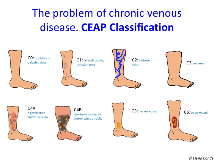
Chronic venous insufficiency, by definition, is associated with skin alterations. While we use the term chronic venous disease to refer to the whole spectrum of morphological or functional venous alterations, with their respective signs and symptoms, the term venous insufficiency is limited to the most advanced stages, in which significant oedema, skin changes or ulceration have already developed. If we use the CEAP classification, chronic venous disease includes all stages (C0-C6) and chronic venous insufficiency only includes C3-C6.

In fact, eczema secondary to venous hypertension and lipodermatosclerosis are the characteristic findings of stages C4a and C4b, respectively.

Eczema secondary to venous hypertension, also known as stasis dermatitis, is characterized by erythema and desquamation, often accompanied by erosions and scabs. Itching is variable, but usually intense. This chronic dermatitis may have episodes of acute exacerbation, usually associated with poor control of venous hypertension, with the development of more lesions, usually more erythematous and exudative.

Eczematous lesions in the lower limbs may spread to other locations, such as the trunk or upper extremities. Over time, this chronic inflammation causes the skin lichenification (thickening) and warty papules in a cobblestone pattern arise.

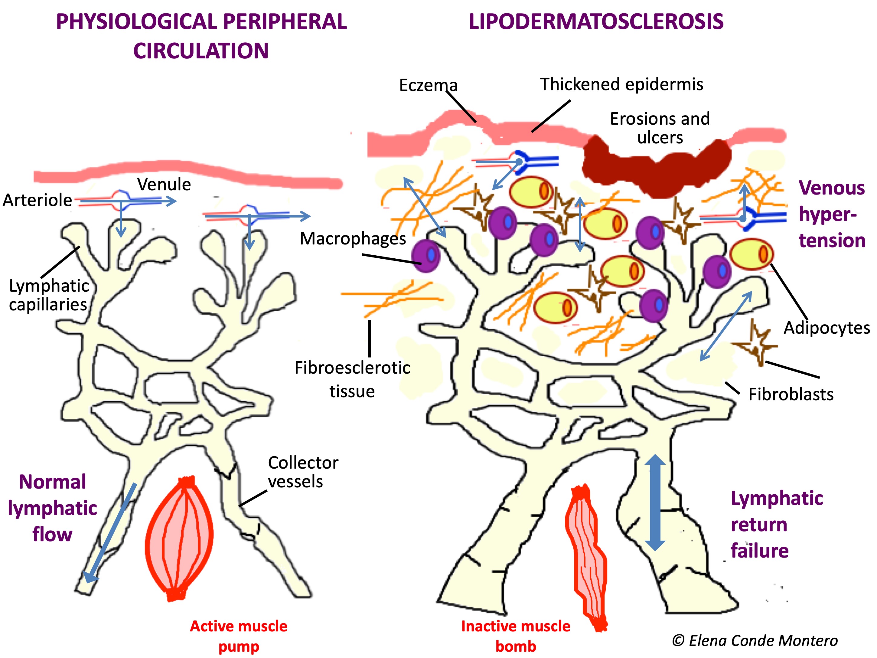
When inflammation secondary to venous hypertension is maintained over time, pericapillary fibrin cuffs and proliferation of fibroblasts in the dermis make it difficult to exchange nutrients. Consequently, the skin and subcutaneous cellular tissue lose elasticity and their consistency becomes woody. These changes are called lipodermatosclerosis. The typical morphological alteration of the legs in these patients is  known as “sign of inverted champagne bottle”.


In the acute phase, lipodermatosclerosis can present with very painful erythematous plaques and can be misdiagnosed as cellulitis, phlebitis or other panniculitis. It is frequent that this induration and progressive fibrosis is associated with secondary lymphedema, by failure of the lymphatic return, involving the dorsum of feet and toes.

The alteration of the immune microenvironment due to damage to the cutaneous and lymphatic integrity (the skin and lymphatic system are key to the immune system) implies an increased risk of infection.


Prevention and treatment of these skin alterations is essential not only to prevent the development of venous ulcers, but also to improve the quality of life of our patients and their perception of their disease severity.

PREVENTION AND TREATMENT OF ALL INFLAMMATORY ALTERATIONS SECONDARY TO VENOUS HYPERTENSION IS BASED ON THE CONTROL OF THAT VENOUS HYPERTENSION. THERAPEUTIC COMPRESSION THE ANTIHYPERTENSIVE AND ANTI-INFLAMMATORY TREATMENT FOR VENOUS HYPERTENSION.

And what else can we do to reduce skin inflammation in these legs?

Stasis eczema, like all inflammatory dermatoses, benefits from topical corticosteroid treatment.

How often should topical corticosteroid be applied and how long should the treatment last? Which corticosteroid should I use? You will not find the answer to these questions in any guideline. What is widely accepted is that adequate control of skin inflammation is essential in order to avoid chronification of the lesions and the consequent risk of wound and infection occurrence. The treatment of the acute outbreak of eczema or lipodermatosclerosis is just as important as maintenance treatment during the chronic phase to avoid exacerbations. In our wound clinic we commonly use medium-high potency corticosteroids in cream. In the acute phase, the application would be daily if the patient wears compression stockings (at night, when the stocking is removed) and 2-3 times a week if the patient wears a bandage. Once this phase has been controlled, after approximately 2 weeks (the duration varies depending on the extent of the lesions), we would move on to maintenance treatment, with daily hydration. If exacerbations are frequent, despite adequate therapeutic compression, we recommend the application of topical corticoid 1-2 times a week, for months, to control subclinical inflammation.


Which dressing should I choose to cover the skin graft donor site?

“How do you cover the donor site? This is a question that always arises when I talk about the punch grafting technique that we do in our clinic. And it is an essential question, since we have to optimize the care of that new wound that we have generated in order to heal another one. Our priorities are to avoid pain, control bleeding, exudate and to favour a fast complete epithelialization.

The idea of writing this post came to me when I read a recent review of dressings used in the donor site.1

Before continuing, as a reminder, the most widespread type of partial thickness skin graft is the mesh graft obtained with dermatome. However, in our practice, where we treat predominantly leg wounds, we use punch grafting (See post: Types of grafts to cover chronic wounds: which one should you choose). The grafts can be obtained with a punch, scalpel blade or curette.

The bleeding in the three cases will be point-sized because we reach papillary dermis (see post: “Differences between epidermal grafts and dermo-epidermal punch grafts).  The presence of multiple free nerve endings in the dermo-epidermal junction, which are exposed or damaged after obtaining the thin skin graft, explains pain in the donor site.

Despite the different techniques for obtaining thin split-thickness skin grafts, the recommendations for management of the donor site are valid for all of them.

Since there is no consensus and, depending on the wound clinic, the protocol of care of the donor site may vary, let’s first see what the studies say. Later, I will tell you what we do in our wound clinic.

Let’s go back to the review1 that was designed to answer the interesting question: which dressing is most effective in reducing pain and accelerating healing of the graft donor site? Thirty-five articles (from clinical trials to case series) were included in the review. All studies used pain scales, but the type of scale and timing of measurement was highly variable. None of the studies detailed the analgesic drugs used, an essential variable in this type of studies. The definition of complete healing also varied between studies. Returning to the objective question of the review, and taking into account the limitations of the study in making comparisons, no dressing has shown superiority. However, interesting conclusions can be drawn from this review:

  • Most papers comparing dry healing with the use of dressings designed to maintain a moist environment in the wound bed, find better pain control and faster healing in the second group.
  • Although a dressing may provide a higher pain reduction in the first few days after the procedure, its power of absorption and the consequent frequency of dressing changes required until complete epithelialisation must be taken into account. For example, a clinical trial2 comparing polyurethane film with alginate underlines that, although the former shows a greater initial analgesic effect, haemostatic power and exudate management with fewer dressing changes makes alginate an interesting alternative, even though there are no significant differences in time to complete epithelialization (21.9 days in the polyurethane film group vs. 18.8 in the alginate group).
  • The secondary dressing is also important to regulate the wound microenvironment and is not usually detailed in the studies.
  • As the healing process evolves, it would be interesting to change the type of dressing, depending on the needs of the wound and the patient, always prioritizing comfort.

“Rapid epithelialization and pain control” should be our goal with the donor site. Before focusing on our experience, I am going to comment on a very interesting strategy that has just been published: mincing the remains of the laminar graft into very small fragments (wounds are irregular and there is usually excess tissue from the graft), uniformly spreading them on the entire surface of the donor site, prior to covering it with a calcium alginate dressing.3 The authors present a clinical trial including 96 patients. An average reduction of 4 days was found until complete epithelialisation (9.1 days in the treatment group and 13.1 days in the control), regardless of the size of the donor site. The benefit in epithelialization may be associated with keratinocytes, growth factors and other cytokines released onto the wound bed. In fact, this is the behaviour that may also have the punch grafts that are observed to accelerate healing despite not achieving appropriate graft taking in the wound bed. Undoubtedly, the excellent proposal of these authors is a very efficient way not to waste tissue!

And now, welcome to our wound clinic! This is the operating table with the material necessary to obtain the grafts (in this case with scalpel blade) and cover the donor site. We use calcium alginate dressing as a primary dressing (which we usually apply in several layers) and gauze with adhesive as a secondary dressing. We always try to place the dressings under local pressure.

Sometimes, in the first dressing changes, which we always try to space out as much as possible, patients refer pruritus in the donor site. When the alginate dressing is removed, always carefully (if it is stuck we moisten it to get an atraumatic detachment), we may find bright erythema and oedema limited to the donor site.

This inflammatory reaction is managed by increasing the frequency of dressing changes and applying topical corticosteroid cream. If this reaction arises early and the wound bed is  exudative and still needs alginate coverage, in each dressing changes we apply gauzes soaked with a corticosteroid solution, and leave them on the wound bed for some minutes. When the majority of wounds are in the final scab phase, the patient can apply a moderate potency corticoid cream at night for 5-7 days, without the need for dressing coverage.

In a preventive way, since its appearance is more frequent when we perform grafts in large areas or if the patient has presented this reaction in previous procedures, we apply gauzes soaked with the corticoid solution immediately after obtaining the grafts.

If there is no more bleeding and the wounds are covered by scab, there are different options to accelerate epithelialization and reduce discomfort in the area. One of them is to apply repairing creams with hyaluronic acid (see post Reasons for the hyaluronic acid boom in wound healing), zinc (see post Why we use topical zinc in wounds and perilesional skin) or other pro-cicatrizing and anti-inflammatory active principles until complete epithelialization (normally in 2-3 weeks), at which time the patient would begin to apply his usual emollient cream.

This is an example of the typical clinical appearance of the donor site on the day of the procedure, at 2 weeks, 3 and 6 months later. The scar is usually not palpable and its final coloration depends on the phototype, the age of the patient and the duration of the inflammatory process until healing (the shorter, the better aesthetic results).

On many occasions we graft wound beds that are not optimal, so we may need to repeat the procedure several times. This is not a problem for patients, since punch grafting is a very well accepted and tolerated technique, which we perform in the consultation room. This is an example of a donor site from which grafts have just been obtained, adjacent to a scar area where the procedure had been performed a few months earlier.


This is our experience. What coverage of the donor site do you prefer?


References:

1.   Brown JE, Holloway SL. An evidence-based review of split-thickness skin graft  donor site dressings. Int Wound J. 2018 Aug 17.

2.   Läuchli S, Hafner J, Ostheeren S, Mayer D, Barysch MJ, French LE. Management of split-thickness skin graft donor sites: a randomized controlled trial of calcium alginate versus polyurethane film dressing. Dermatology. 2013;227(4):361-6. 

3.   Miyanaga T, Haseda Y, Sakagami A. Minced skin grafting for promoting epithelialization of the donor site after split-thickness skin grafting. Burns. 2017 Jun;43(4):819-823.


Why do calcifications occur in chronic skin ulcers?

Since this is not an unusual finding, but there is little scientific literature on it, I will dedicate today’s post to dystrophic calcifications in chronic venous ulcers.

When we speak of calcifications we are referring to abnormal deposits of insoluble calcium salts. When they are arranged in an organized way forming bone, they are called ossifications.

Calcinosis cutis is the term used when this calcification occurs in the skin tissue and can be classified as follows (Urbina, 2001):

  • Dystrophic calcification, due to tissue damage secondary to trauma, burns and inflammatory, degenerative or  neoplastic diseases.
  • Metastatic calcification, associated with various disorders that occur with elevated plasma levels of calcium and /or phosphorus.
  • Idiopathic calcification, which is not related to tissue damage or metabolic disorders.

The most frequent group is dystrophic calcifications. Among the inflammatory causes is chronic venous insufficiency, with or without the presence of a cutaneous ulcer. Calcium-phosphorus metabolism and serum levels are normal. Different hypotheses have been proposed to explain the development of these ectopic calcifications. Anoxia and inflammation that occur in the legs with venous hypertension involves cell necrosis and the release of cells and inflammatory mediators, with the production of free radicals and damage to connective tissue (collagen, elastin) or subcutaneous fat. In this damaged tissue, intracellular calcium levels may increase, with the consequent formation and precipitation of crystals. On the other hand, denatured proteins bind to phosphate and allow them to bind to calcium, with consequent precipitation of calcium phosphate.

The diagnosis can be made by palpation and visualization in radiography or ultrasound (Piry, 1992).

When the patient presents a cutaneous ulcer, this calcification can become visible, as a hard material of bone consistency and dark brown coloration. Normally they are strongly adhered to the wound bed, with significant pain when trying to pull on them. These deposits act as a foreign body, which promotes the inflammatory response in the wound and therefore slows healing. A retrospective study of 212 patients with chronic leg ulcers (no response to conventional treatment for at least 3 months) found calcifications in 18% and ossifications in 7% of patients. The presence of these abnormal deposits was associated with greater pain and worse response to compressive treatment (Wollina, 2009).

The main radiological differential diagnosis has to be made with phlebolites, which correspond to parietal venous calcifications after episodes of deep vein thrombosis.

There are different options in the treatment of these ossifications in skin ulcers, but all involve their removal from the wound bed (Chaverri, 2011; Enoch, 2005):

  • Achieve detachment of calcification by moisturizing the wound bed with hydrogel-type dressings
  • Mechanical removal with tweezers and scalpel. Since it is painful, it is recommended to previously anaesthetize the area.
  • Surgical excision under anaesthesia
  • Radical debridement of the ulcer, with or without subsequent coverage with skin graft.

Referencias:

  1. Chaverri Fierro D, Portas Freixes J, Bosch Cuenca M, Sáez Horts M, Montoya Hellín I, González Sierra M et al. Calcificación distrófica en úlceras de la extremidad inferior. Gerokomos 2011; 22 (3): 126-130.
  2. Enoch S. Kupitz S, Miller DR and Harding KG. Dystrophic calcification as a cause for non healing leg ulcers. Int Wound J 2005; 2:142-147.
  3. Piry A, Vin F, Allaert FA. Inflammation and subcutaneous calcification of venous origins. Fhlebologie 1992; 45 (1): 41-7.  
  4. Urbina F, Pérez L, Sudy E, Misad C. Calcificación y osificación cutánea. Actas Dermosifiliogr. 2001;92:255-69.
  5. Wollina U, Hassenöhrl K, Köstler E, Schönlebe J, Heinig B, Haroske G, Kittner T, Dystrophic calcification in chronic leg ulcers – a clinocopathologic study. Dermatol Surg 2009; 35(3): 457-61.


Ulcer secondary to alteration of homocysteine metabolism mimicking pyoderma gangrenosum

Homocysteine is an amino acid that is not present in dietary proteins, but is formed exclusively as an intermediate product in the metabolism of methionine to cysteine and participates in the folate cycle and DNA methylation. If there is any mutation in the enzymes involved or there is an acquired deficit of essential vitamins in these metabolic processes, homocysteine blood levels increase.

High blood levels of homocysteine (hyperhomocysteinemia) are considered a risk factor for arterial and venous thrombosis.

Vascular alterations produced by homocysteine are thought to be multifactorial. Homocysteine causes direct damage to the endothelium, stimulates lipid peroxidation and increases platelet aggregation. In addition, this amino acid promotes inflammation in vessels by releasing pro-inflammatory cytokines and stimulating leukocyte adhesion to endothelial cells, with consequent migration to surrounding tissue.1

As we can observe in the following scheme, the activity of the enzyme methylenetrahydropholate reductase (MTHFR), involved in the folate cycle, is essential for the reconversion of homocysteine into methionine. The polymorphisms of this enzyme can decrease its action and, therefore, increase the concentration of homocysteine in the blood. The C677T mutation (mutation in a single nucleotide) is the most frequent polymorphism and is an independent risk factor for thrombosis, even with normal homocysteine levels in the blood. In patients with coronary artery disease or hypertension its prevalence increases to 25-35%. Risk increases when cellular folate levels decrease.2 Thrombophilia and vasculopathy associated with hyperhomocysteinemia may be the trigger for skin wounds.3


Higher serum homocysteine levels have been detected in patients with chronic venous insufficiency (with or without ulcer) than in patients without this underlying disease. Therefore, elevation of serum homocysteine may be implicated in the pathogenesis of stasis dermatitis and its progression to ulceration.1 Another study has shown the benefit in healing of daily folic acid supplementation in cases of venous ulcer and hypermicysteinemia.4

Regardless of their possible role in chronic venous insufficiency, cases of pyoderma gangrenosum-like necrotizing lesions caused by disorders in homocysteine metabolism have also been reported.2

Diagnosis of pyoderma gangrenosum is a diagnosis of exclusion. Clinically it can be confused with different diseases. Normally we have in mind the most frequent differential diagnoses: vasculitis, occlusive vasculopathy, infections, malignancies, Martorell ulcer, even factitious dermatitis. It is important that the skin biopsy is sufficiently broad and deep to exclude these diagnoses, as many share the typically neutrophilic inflammation that can be seen histologically in pyoderma gangrenosum.

However, the diagnostic key is not always provided by biopsy, as in the case of alterations in homocysteine metabolism. It is important to know this pathology in order to suspect it in patients with these diagnoses who do not respond as expected to treatment.


Right lateral leg ulcer and necrotic scabs with erythemato-violaceous halo on the left lateral leg in the context of hyperhomocysteine (lesions previously diagnosed as pyoderma gangrenosum without response to immunosuppressive treatment).


We need to request homocysteine blood levels. However, it is important to know that, despite normal homocysteine levels, a patient may have a mutation in the MTHFR enzyme and therefore an alteration in the metabolism of this amino acid with clinical repercussions. 2 In this case, with clinical suspicion, despite normal homocysteine levels, mutation in the MTHFR gene must be requested. Treatment with vitamin B6, vitamin B12 and folic acid supplementation may reduce homocysteine levels, even in patients with normal serum concentration of these vitamins.2,3

Undoubtedly, considering the simplicity and good tolerance of treatment, it is very interesting to go deepler into the potential role of alterations in homocysteine metabolism in recalcitrant ulcers in patients with risk factors and history of cardiovascular events.

Have you diagnosed any cases of ulcers in the context of hyperhomocysteinemia / MTHFR mutation?


Referencias

1.    Durmazlar SP, Akgul A, Eskioglu F. Hyperhomocysteinemia in patients with stasis dermatitis and ulcer: a novel finding with important therapeutic implications. J Dermatolog Treat. 2009;20(6):336-9. 

2.    Rubio-González B, Castellanos-González M, Alegría-Landa V, del Mar Burgués-Calderón M, Zarco-Olivo C, Vanaclocha-Sebastián F. Skin ulcers mimicking  pyoderma gangrenosum in a patient with MTHFR polymorphism. J Am Acad Dermatol. 2012 Dec;67(6):e282-4. 

3.    New D, Eaton P, Knable A, Callen JP. The use of B vitamins for cutaneous ulcerations mimicking pyoderma gangrenosum in patients with MTHFR polymorphism. Arch Dermatol. 2011 Apr;147(4):450-3. 

4. de Franciscis S, De Sarro G, Longo P, Buffone G, Molinari V, Stillitano DM, Gallelli L, Serra R. Hyperhomocysteinaemia and chronic venous ulcers. Int Wound J. 2015 Feb;12(1):22-6