Home Blog Page 10

Suspected infection in a leg ulcer: swab or punch biopsy?

We have a patient with a venous ulcer, for severeal months, which has been stagnant for two weeks. Exudate has increased despite adequate compressive therapy and it is viscous, greenish and has a worse odor than in previous dressing changes. Wound edges are erythematous and warm. The patient reports more pain than usual and has a distermal sensation, without any other associated systemic symptoms. These clinical signs and symptoms suggest infection, so we will take a sample for microbiological culture.

Watch out! The microbiological study helps us identify the bacteria involved, but the diagnosis of infection is clinical. Therefore, the order of action will always be as follows: 1st suspicion of infection during the clinical examination, 2nd obtaining a sample for culture. In summary, this seems very simple, but these two points are a source of controversy, not to mention the selection of treatment, both topical and systemic!

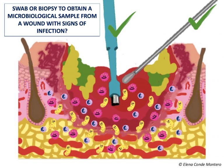
In this post we are going to focus on the eternal doubt when selecting the way to collect the sample: swab or punch biopsy?

I’m going to focus on leg wounds, which are the tyoe of wound that we treat most often in the clinical practice. In my clinic, in the presence of infection criteria in a leg ulcer, we usually collect the sample with a swab. However, in the absence of response to the antibiotic prescribed by the antibiogram, we perform a biopsy. In addition, if the torpid evolution of a leg ulcer makes us reconsider its etiology and we will perform a biopsy for histological study, if it meets the criteria for infection, we also send a tissue sample for microbiological study. In case of suspected infection by fungi or mycobacteria, we make sure that the biopsy is processed properly.

Now we will see what the studies and guidelines say, but first I will point out some general principles and summarize the characteristics of each technique.

General principles of collecting samples for culture:

  • In a chronic wound with no signs of infection, bacterial contamination and colonising bacteria are found, which normally are not preventing healing. Therefore, since these bacteria do not require treatment, they do not need to be routinely sampled for identification.
  • Although surface samples may not be an accurate reflection of the microbiological environment of the tissue at depth, any bacteria present in deep layers are very likely to be at the surface as well.1
  • The amount of bacteria detected in a quantitative analysis of the sample cannot be considered as an isolated criterion for infection, since its clinical impact depends on the bacterial species and the patient’s immune response.
  • Before sampling, the wound should be cleaned with saline and any devitalised tissue removed.
  • In addition to wound swab and biopsy, there are other techniques, such as needle aspiration of the wound edge.
  • The most frequently isolated bacteria in leg wounds with suspected infection is aureus, but it is usually polymicrobial flora.
  • A proper protocol for processing the sample must be followed.
  • Each laboratory will choose the microbiological techniques to be applied to each sample according to its availability.


Wound swab:

  • This is the most practical and simple alternative. Some experts consider that, as it is a superficial sample, it can make it difficult to differentiate between contamination and infection.
  • There are different techniques for taking the sample, but the most classic are the Levine technique (pressure in a 1 cm2 area of the wound bed for 5 seconds) and the zig-zag technique (displacement with rotation of the swab by 10 points through the entire wound bed, without touching slough or the edges of the lesion). Levine technique has shown greater reliability, having greater microbiological concordance with biopsy samples.2
  • It allows a semi-quantitative study, which is easier to perform than quantitative studies


Punch biopsy:

  • Tissue biopsy is a deep sample and it is the gold standard, that is, it is considered the most sensitive and specific test to detect the bacteria involved in the infection. However, its obtaining requires local anesthesia and, as it is an invasive procedure, it is not possible in all clinical settings.
  • A fragment of tissue is removed from the wound bed, avoiding sloughy and necrotic tissue.
  • The punch is a tool that facilitates its collection (usually we use 4 mm), but you can use a scalpel blade or a curette.
  • After it has been carried out, the placement of alginate and pressure at the point where the sample has been taken promote haemostasia.


What do the studies say?

Studies are scarce, with few patients, and most of them do not specify the swabbing technique used. The correlation between the two techniques varies from non-existent to high. It is difficult to compare the results of studies with wounds of different aetiologies (diabetic foot, pressure ulcers, burns),3 so I will focus on leg wounds. I will highlight the study with the most favourable results for swabbing: “No more need for biopsies”.4 It included 46 patients with hard-to -heal venous ulcers. All wounds were cleaned with serum but not debrided. When comparing the bacterial species isolated with biopsy (punch 4 mm) and with swab (no method of collection specified), no significant differences were found. In order of prevalence, the most frequently isolated species in this Danish study were S. Aureus, E. Faecalis and Ps. Aeruginosa. This study also finds that the greater the number of samples collected, regardless of the technique, the greater the probability of finding a greater number of species. This is explained by the polymicrobial environment of chronic wounds, with a specific distribution of bacteria in the wound. Wound swabs, which may contact a larger area of the wound than the biopsy, might better reflect the microbiological environment of the wound. The authors conclude that the swab is an interesting technique for the microbiological study of infected wounds in our usual clinical practice and, after good cleansing and debridement, the superficial bacterial load corresponds to that in deep tissues.

A recent review5 on samples for culture in infected wounds highlights that, although the swab is a good option in the initial evaluation, in case antibiotic treatment does not obtain a clinical response, the biopsy test is interesting.

A conclusión that is repeated in the studies is that the microbiological result depends not only on the method used to collect the sample, but also on its adequate processing, including the type of swab material, the transport medium, the time to analysis and the interpretation criteria (qualitative, semi-quantitative or quantitative). The review “Microbiological diagnosis of skin and soft tissue infections “1 clearly explains the phases of sample collection, processing and interpretation.


What do the guidelines say?

To answer this question I have turned to the document published by the EWMA (European Wound Management Association) in 2016 “Management of patients with venous leg ulcers. Challenges and current best practice”, which reviews the recommendations of 8 clinical guidelines for the management of venous ulcers. The document emphasizes that diagnosis of infection is clinical, that all chronic wounds are colonized by bacteria and that sampling for culture is only of interest if there are signs and symptoms of infection. The recommendations on the technique for obtaining the sample include swab and biopsy, without detailing or mentioning the superiority of either.

When reviewing practical guidelines, with summary recommendations on how to perform the sample collection (such as this British example: Venous leg ulcers: Infection diagnosis and microbiology investigation), swab collection is the most widespread.

Following the recommendations of the SEIMC (Sociedad Española de Enfermedades Infecciosas y Microbiología Clínica), we should take more than one sample from different areas of the wound, since an isolated sample may not detect all the microorganisms causing the infection.

What do you do in your daily clinical practice?


References:

1. Burillo A, Moreno A, Salas C. [Microbiological diagnosis of infections of the  skin and soft tissues]. Enferm Infecc Microbiol Clin. 2007 Nov;25(9):579-86.

2. Gardner SE, Frantz RA, Saltzman CL, Hillis SL, Park H, Scherubel M. Diagnostic validity of three swab techniques for identifying chronic wound infection. Wound  Repair Regen. 2006 Sep-Oct;14(5):548-57.

3. Nelson A, Wright-Hughes A, Backhouse MR, Lipsky BA, Nixon J, Bhogal MS, Reynolds C, Brown S; CODIFI collaborators. CODIFI (Concordance in Diabetic Foot Ulcer Infection): a cross-sectional study of wound swab versus tissue sampling in infected diabetic foot ulcers in England. BMJ Open. 2018 Jan 31;8(1):e019437.

4. Gjødsbøl K, Skindersoe ME, Christensen JJ, Karlsmark T, Jørgensen B, Jensen AM, Klein BM, Sonnested MK, Krogfelt KA. No need for biopsies: comparison of three sample techniques for wound microbiota determination. Int Wound J. 2012 Jun;9(3):295-302.

5. Copeland-Halperin LR, Kaminsky AJ, Bluefeld N, Miraliakbari R. Sample procurement for cultures of infected wounds: a systematic review. J Wound Care. 2016 Apr;25(4):S4-6, S8-10.


Does it make sense to apply topical corticosteroids to improve the healing of superficial burns?

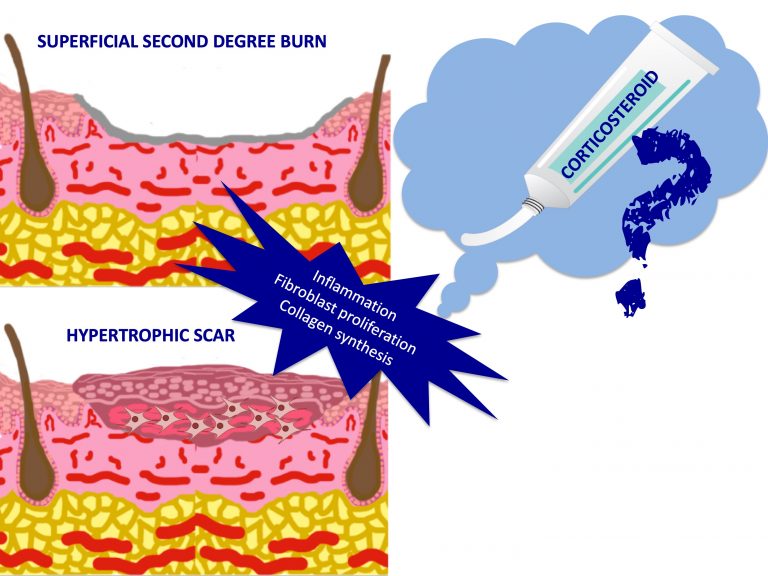
A burn is a very common type of traumatic wound, whose healing process can be pathological, with the appearance of a hypertrophic or keloid scar. Intralesional corticosteroid infiltration is a technique of choice for treating pathological scars. Therefore, considering the known risk of hypertrophic scarring after a burn, could it be interesting to apply topical corticoid in a preventive manner? The authors of an article published in 20141 ask this same question and conclude that ” Theoretically, topical corticosteroids could reduce pathological scarring by decreasing inflammation and activation of fibroblasts. However, there are no studies evaluating this hypothesis”.

Despite the absence of studies on this subject, we will understand the reason of this interesting reflection. To this end, we begin with a brief introduction to burns.


How are burns classified?

Depending on the depth of skin involvement, burns are classified in different degrees:

  • First degree: only the epidermis is affected. Usually only erythema occurs and heals without leaving a scar.
  • Second degree: affects epidermis and dermis with the formation of a blister. Depending on the depth of the affection of the dermis, they are divided into superficial and deep, normally the bottom of the blister being red in the former and whitish in the latter. The risk of developing a hypertrophic or keloid scar is greater the deeper the involvement.
  • Third degree: it implies a necrosis of all the skin layers. Graft coverage must be performed to avoid contracture and hypertrophy.

In addition to the depth of the affectation, to measure its severity, its extension, location, type of burn, age and associated pathologies of the patient must be taken into account.

We will now focus on a type of burn that we frequently encounter in our daily lives, superficial second-degree burns.

What is the healing process after a burn?

As in any wound, after the damage produced by a burn, a process begins in which the cells and molecules involved vary over time. Therefore, to facilitate its study, the healing process is divided into phases, although these phases actually overlap (see post “Brief overview of wound healing”)

In the inflammatory phase, macrophages are first responsible for creating a pro-inflammatory environment, releasing cytokines such as IL-1 and TNF, and they engage in phagocytosing bacteria and destroying devitalised tissue. Later, in the proliferative phase, macrophages differentiate to secrete growth factors (TGFb1 and 2, PDGF) that stimulate the proliferation of fibroblasts and their production of collagen (type III collagen), as well as the proliferation of endothelial cells for the formation of new vessels. In the final or remodeling phase, apoptosis of the cells formed for the proliferation of new wound tissue occurs and type III collagen is degraded to form type I collagen. This collagen is organized differently than normal skin collagen, but over time the appearance and function of the scar can be improved.

If excessive scar tissue has been produced, a hypertrophic scar (red, raised, that worsens months after wound production and improves spontaneously over the next 1-2 years) or a keloid (extends beyond the limits of the original wound and does not improve without treatment) may develop.

With respect to other types of wounds, burns are injuries with a high risk of pathological healing, mainly if there is deep dermis involvement. This higher incidence of hypertrophic or keloid scars in burns seems to be associated with the intense and prolonged inflammation that usually occurs in this type of wounds and the consequent excessive proliferation of fibroblasts and collagen production.


Why and how should topical corticosteroids be applied to prevent pathological scarring?

Topical corticosteroids decrease the synthesis and release of inflammatory mediators (including prostaglandins and cytokines such as IL-1 and TNF), reduce the release of growth factors that stimulate fibroblast proliferation (TGFb1, TGFb2, PDGF) and thus decrease collagen production. Thus explained, it seems an interesting preventive strategy to prevent a pathological scar in a superficial burn. However, when to start? and how long to maintain it? These are difficult questions to answer, essentially because scar hypertrophy can occur months after the burn. In fact, the authors of the article with which I started this post,1 propose the use of topical corticosteroids after epithelialization to limit possible persistent inflammation in the dermis, which would be responsible for excess fibroblasts and collagen causing hypertrophy.

The few studies that have been published on topical corticosteroids in first and second degree superficial burns do not help us to answer these questions, as they focus on its analgesic and early anti-inflammatory effect. Some authors propose the benefit of its early and time-limited application (a few days) to decrease inflammation and pain without affecting subsequent epithelialization. However, two clinical trials with healthy volunteers (15 and 20 volunteers included) found no improvement with respect to placebo when applying medium or high potency corticosteroids in the first 24 hours2 or daily for the first 3 days after the burn3.


And what do the guidelines say about topical corticosteroids in superficial burns?

The guidelines produced by the Japanese Dermatology Association,4 published in 2016, make recommendations on the use of topical corticosteroids in first- and second-degree superficial burns. It is stressed that its use in this indication is based on its anti-inflammatory activity and that, despite the lack of available evidence, it is a widely used treatment in clinical practice and recommended by experts.

What is your experience? Do you recommend medium or high potency topical corticosteroid cream for superficial second-degree burns? Early or after epithelialization?


Referencias:

1.  Taheri A, Mansoori P, Al-Dabagh A, Feldman SR. Are corticosteroids effective for prevention of scar formation after second-degree skin burn? J Dermatolog Treat. 2014 Aug;25(4):360-2

2.  Faurschou A, Wulf HC. Topical corticosteroids in the treatment of acute sunburn: a randomized, double-blind clinical trial. Arch Dermatol. 2008 May;144(5):620-4. 

3.  Pedersen JL, Møiniche S, Kehlet H. Topical glucocorticoid has no antinociceptive or anti-inflammatory effect in thermal injury. Br J Anaesth. 1994 Apr;72(4):379-82 

4.  Yoshino Y, Ohtsuka M, Kawaguchi M, Sakai K, Hashimoto A, Hayashi M et al. The wound/burn guidelines – 6: Guidelines for the management of burns. J Dermatol. 2016 Sep;43(9):989-1010. 

Martorell Ulcer and Vitamin K Antagonists: A Dangerous Combination

This week, two scientific meetings that I usually attend every year, have taken place: the 46th National Congress of Dermatology and Venereology in Palma and the EWMA 2018 Conference in Krakow.

It was a pleasure to chair a session about wound healing at the Spanish National Congress of Dermatology, where we talked about diagnostic and therapeutic pearls in the treatment of chronic leg ulcers.


This post is based on a pearl presented by my colleague and best friend Celia Horcajada: “The replacement of acenocoumarol by another oral anticoagulant in patients with Martorell ulcer”. Curiously, that same day, in Krakow, Jürg Hafner gave a talk in which this pearl had to be mentioned as well.


Martorell ulcer, as proposed by Dr. Jürg Hafner in his excellent article Calciphylaxis and Martorell Hypertensive Ischemic Leg Ulcer: Same Pattern – One Pathophysiology,1 is a hypertensive ischemic arteriosclerosis that can be framed within the clinical-physiological spectrum of calciphylaxis. In fact, both entities share, as we can see in the table, risk factors, clinical and histological aspects.

This concept is relevant to better understand the physiopathology of these two entities and to propose a therapeutic approach with common strategies, such as debridement, negative pressure therapy, early grafting or sodium thiosulfate. Furthermore, in both Martorell ulcer and calciphylaxis it is essential to control cardiovascular risk factors and assess the replacement of the vitamin K antagonist by low molecular weight heparin or factor Xa inhibitors.


And why is it interesting to withdraw acenocoumarol in these patients?

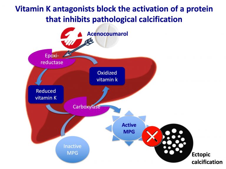
Acenocoumarol has an anticoagulant action because it inhibits the activation of coagulation proteins that depend on vitamin K (prothrombin, factor VII, IX, X). Because of this dependence on vitamin K, these coagulation factors are part of a family of proteins called G-proteins. This vitamin K-dependent protein group also includes anticoagulant factors, such as vitamin S or C (so in patients with a deficit of these factors, skin necrosis can occur due to acenocoumarol) and the calcification-inhibiting protein complex Fetuin A (or glycoprotein α2-Heremans-Schmid)-GLA-matrix protein (or MGP). In order for the binding and subsequent activation of this protein system to take place (as with all other Gla-proteins), the glutamic residues of the GLA-matrix protein have to undergo vitamin K-dependent carboxilation. For this carboxilation to be possible, the vitamin K has to be converted to its reduced form by an epoxide-reductase enzyme.

Acenocoumarol blocks the action of the epoxide-reductase, so it inhibits the activation of this transport protein complex responsible for binding insoluble calcium phosphate and inhibiting pathological calcification.

These proteins were discovered in the 1990s and their existence explained the nephrologists’ hypothesis that vitamin K antagonists could worsen cases of calciphylaxis. The way this calcification inhibitor complex acts is not completely clear, but its role seems key to prevent ectopic calcification in patients with predisposing pathologies. Vitamin K deficiency and the consequent decrease in the concentration of carboxylating GLA matrix protein have been associated with an increased risk of calciphylaxis.2


Therefore, in patients being treated with acenocoumarol in whom we suspect lesions in the ulcer spectrum of Martorell-calciphylaxis, replacement with another anticoagulant (low molecular weight heparin or activated factor X inhibitors) is recommended.


References:

  1. Hafner J. Calciphylaxis and Martorell Hypertensive Ischemic Leg Ulcer: Same Pattern – One Pathophysiology. Dermatology. 2016;232(5):523-533.
  2. Nigwekar SU, Bloch DB, Nazarian RM, Vermeer C, Booth SL, Xu D, Thadhani RI, Malhotra R. Vitamin K-Dependent Carboxylation of Matrix Gla Protein Influences the Risk of Calciphylaxis. J Am Soc Nephrol. 2017 Jun;28(6):1717-1722. 

Sulodexide: a pill against endothelial damage from venous hypertension

The idea to write this post came after reading the scientific program of the 13th Wound Specialist Day in Zürich. I find this year’s theme very interesting “When an ulcer is healed: prevention of recurrence”. One of the talks is dedicated to systemic treatment, a topic for which I am developing a growing interest.

If we think about the treatment of a venous ulcer, the alternatives that probably come to mind are compression therapy, dressings, skin grafting and surgery or endovenous procedures. And why do we forget about pharmacological treatment, perhaps because its benefit is not clear?

In the post “Ulcers and post-thrombotic syndrome: a therapeutic challenge” I referred to the interest of pharmacological treatment to protect the endothelium. We cannot forget that a venous ulcer is a consequence of capillary and subcutaneous cellular tissue damage produced by venous hypertension and, therefore, any action that decreases the damage at that level is interesting. Regardless of the underlying cause of venous hypertension, an anti-inflammatory strategy must be established to reduce the damage that is occurring in the endothelium due to the release of free radicals, pro-inflammatory cytokines and an imbalance between metalloproteinases and their inhibitors. The interaction between activated leukocytes and the damaged endothelium promotes thrombosis and the migration of inflammatory cells into the extracellular matrix, with consequent hypoxia and tissue destruction.1

It would be ideal to be able to help stop this destructive cascade with a pill, wouldn’t it? Let’s see what has been studied about this.

Drugs with antithrombotic and anti-inflammatory properties for the endothelium include pentoxifylline, flavonoids, acetylsalicylic acid and sulodexide. Pentoxifylline is the drug whose utility, in association with compression therapy, has the most evidence at present, with favourable results in the Cochrane review published in 2012. It is an agent with haemorrheological properties, i.e. it improves blood perfusion by making the red blood cell membrane more flexible and encouraging its passage through the vessels, as well as reducing platelet aggregation and fibrinogen levels.1

In this post I will explain in more detail sulodexide, a systemic drug with anticoagulant, antithrombotic and anti-inflammatory properties on the endothelium, which is increasingly present in our clinical practice. It is a mixture of 2 glycosaminoglycans, heparan sulfate (80%) and dermatan sulfate (20%). Unlike heparin, it can also be administered orally and has a lower risk of bleeding. The main novelty in its mechanism of action is its potential protective effect on the endothelium, since it appears to inhibit proteolytic degradation of the glycocalyx (protective layer of endothelial cells that is damaged by shear forces and inflammation secondary to venous hypertension).2


 In addition to its benefit in healing venous ulcers, it has been shown to be useful in preventing the recurrence of venous thromboembolic disease and, because of its protective action on the endothelium, it appears to be interesting from the early stages of chronic venous disease.2

We will focus on its interest in the treatment of venous ulcer, in association with compression therapy and conventional local care. The randomized double-blind clinical trial with the largest sample size is the multicenter study SUAVIS (Sulodexide Arterial Venous Italian Study)3 , which includes 235 patients, divided into a placebo group (conventional treatment) and the treatment group (sulodexide + conventional treatment), with a duration of 3 months. Sulodexide was administered intramuscularly, once a day, for the first 20 days (injection of 600 LRU, Lipoprotein Lipase Realising Units, which are the units in which the dose of Sulodexide is measured). Later, until completing the 3 months of treatment, it was continued with oral administration (500 LRU) 2 times a day. A higher percentage of healed ulcers and a faster reduction of the wound area, statistically significant, were found at month 2 and 3 in the sulodexide treated group. However, a limitation of the study is the high variability between the conventional treatment performed on each patient (no standardization of type of local treatment and medical compression).

This study was analysed together with three other clinical trials, with a total of 463 patients included, in a Cochrane review in 20164. In addition to the heterogeneous sample size of the studies (range 20-235 patients), the dose and route of administration used (oral or intramuscular), as well as the duration of treatment, is variable between the studies (1-3 months). On the other hand, only one study was double-blind3 and conventional treatment was not standardized in 3 of the trials.

No major bleeding events were evident in the studies performed and no significant differences in the adverse events rate were detected between the sulodexide treated group and the control group.

Although a higher percentage of healed wounds, a greater reduction in wound size and an acceleration of healing were found in the sulodexide treated group, the low quality of evidence due to the small number of studies and the limitations of the included trials prevents from drawing conclusions from the actual usefulness of this drug. In addition, studies are needed to measure the impact of sulodexide on recurrence, quality of life and healthcare cost.

What is the real impact of this endothelium-protective strategy for our patients with venous ulcers? What dose and duration of treatment would be most beneficial in promoting healing and preventing relapses? I am sure that new studies will soon answer these questions.


References:
  1. Varatharajan L, Thapar A, Lane T, Munster AB, Davies AH. Pharmacological adjuncts for chronic venous ulcer healing: a systematic review. Phlebology. 2016 Jun;31(5):356-65.
  2. Andreozzi GM. Role of sulodexide in the treatment of CVD. International Angiology 2014;33(3):255–62.
  3. Coccheri S, Scondotto G, Agnelli G, Aloisi D, Palazzini E, Zamboni V; Venous arm of the SUAVIS (Sulodexide Arterial Venous Italian Study) Group. Randomised, double blind, multicentre, placebo controlled study of sulodexide in the treatment of venous leg ulcers. Thromb Haemost. 2002 Jun;87(6):947-52.
  4. Wu B, Lu J, Yang M, Xu T. Sulodexide for treating venous leg ulcers. Cochrane Database Syst Rev. 2016:CD010694.

Sclerotherapy gains against recurrence of venous leg ulcers

Recently, the 45th National Congress of the Spanish Academy of Dermatology took place in Madrid, where, with Dr. Rita Cabeza, I had the honour and pleasure of chairing a session about clinical cases on wound healing. The discussion that followed each presentation was enormously enriching for all. In this session I personally met Dr. Alejandro Sierra, a specialist in Phlebology at the Clínica Universitaria de Navarra. I would like to reflect in this entry the passion he transmitted to me when he told me about the excellent results that have been obtained for years in his Department with sclerotherapy for venous ulcers. For this, he has prepared a presentation with brushstrokes of his experience, which you will find at the end. I would like to thank him and his team, especially Dr. Eugenia Pillado, for the valuable bibliographic and iconographic contributions to this entry.


To put us in the picture, in 2004 the results of the ESCHAR randomized clinical trial were published,1 in which the benefit of superficial venous system surgery combined with compression was compared with compression therapy alone, in patients with active or recently healed venous ulcers (stages C6 and C5 of the CEAP classification, respectively). Although no difference was found in the healing rate at 6 months (65% in the 2 groups), the lowest recurrence rate at one year observed in this study (28% in the compression therapy alone group vs 12% in the compression and surgery groups, p<0.0001) was again found at 4 years of follow-up (56% vs 31%, respectively, p<0.01).2

We cannot forget the patient profile that usually presents this disease, elderly people with many comorbidities. Therefore, minimally invasive endovenous procedures, mainly sclerotherapy, widely used in the treatment of varicose veins, have positioned themselves in recent years as a very interesting alternative to surgery in the management of venous ulcers, with similar results (even better in some cases) and fewer complications.

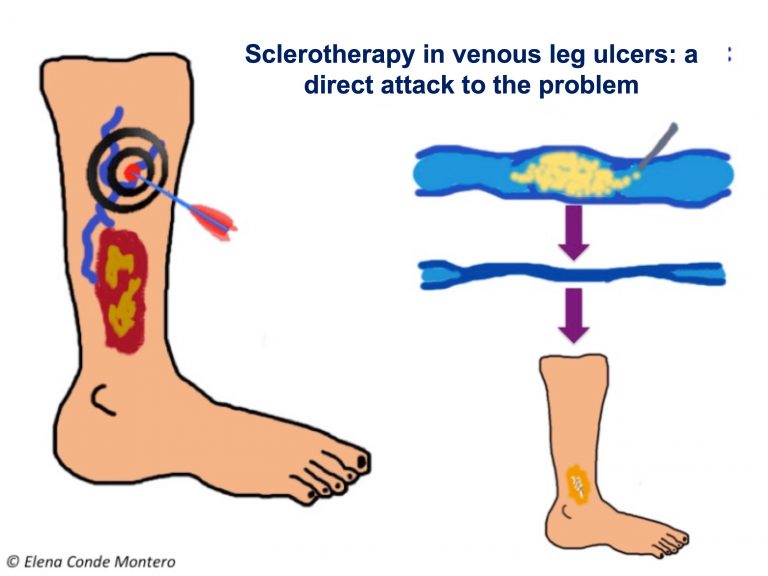
There are different techniques and sclerosing substances. Sclerotherapy with microfoam, which is achieved by vehiculating the liquid sclerosant in microbubbles, is more effective, since it achieves greater contact with the endothelium and, therefore, damage at all points inside the vessel, which it fills completely. When the sclerosing substance is injected into the affected vein, the vein closes due to the endothelial damage and the subsequent fibrosis of its walls. Therefore, the reflux in that path is eliminated. The objective of this treatment, as with surgery or other endovenous techniques (radiofrequency, laser ablation), is to eliminate the incompetent veins that are producing the venous hypertension that causes the ulcer. The special benefit of sclerotherapy, compared to the other alternatives, is the possibility of accessing all the affected veins involved in the reflux, including the smaller ones. The ultrasound control allows to verify that all the incompetent vessels of the superficial venous system have been occluded, which go from the origin or origins of the reflux, pass through its transmission route and reach their destination on the surface (including the smallest vessels of the ulcer bed, those of inframalleolar location and in context of great cutaneous deterioration). This is a fast, safe and cost-effective technique.

The first series of patients with venous ulcers treated with intravenous injection of ultrasound-guided polidocanol microfoam was published by Dr. Cabrera (inventor of the technique for obtaining this sclerosing foam, patented in 1993), in collaboration with the phlebology team of the Clínica Universitaria de Navarra.3 This retrospective series included 116 patients with venous ulcers, in which a healing rate of 83% and a relapse rate of 6.3% at 6 months were achieved.

They have recently published a new series that reflects the excellent results of their experience4 , which are comparable to those obtained by other authors5,6,7.

In this series of 180 patients published by Dr. Pedro Lloret, a healing rate of 79.4% was obtained at 6 months. In the subgroup analysis, the isolated presence of superficial insufficiency (no deep component or incompetence of the perforators) was associated with higher healing rates (95.1%). The recurrence rate at 1, 2 and 3 years was 8.1%, 14.9%, and 20.8%, respectively, with higher risk in patients with perforator vein incompetence or history of surgery. However, the extensive experience of these authors defends that, with adequate follow-up of patients (and periodic sclerosis of refluxes from the superficial venous system, which appear by recanalization of occluded but non-fibrous venous segments, or are generated by new pathways, often from incompetent perforators), the rate of ulcer recurrence can be maintained at less than 4%.


This work also reflects the positive impact of reduced edema and pain control on patient quality of life.


As the previously cited studies show, the benefit of sclerotherapy is not limited to ulcers with isolated superficial venous insufficiency. Its use in ulcers with an arterial component or with affectation of the associated deep venous system, can help to control venous hypertension, with a decrease in edema and with a direct impact on healing.


On the other hand, treatment with sclerotherapy has special interest in patients without adequate adherence to compressive therapy.


Here I leave you with the presentation that the experts of the Clínica Universitaria de Navarra have kindly prepared for us.

  1. Barwell JR, Davies CE, Deacon J, et al. Comparison of surgery and compression with compression alone in chronic venous ulceration (ESCHAR study): randomized controlled trial. Lancet 2004;363:1854– 9 5 .
  2. Gohel MS, Barwell JR, Taylor M, Chant T, Foy C, Earnshaw JJ, et al., Long term results of compression therapy alone versus compression plus surgery in chronic venous ulceration (ESCHAR): randomised controlled trial. BMJ, 2007; 335: 83.
  3. Cabrera J, Redondo P, Becerra A, Garrido C, Cabrera J Jr, García-Olmedo MA, et al. Ultrasound-guided injection of polidocanol microfoam in the management of venous leg ulcers. Arch Dermatol 2004;140:667-73.
  4. Lloret P, Redondo P, Cabrera J, Sierra A. Treatment of venous leg ulcers with ultrasound-guided foam sclerotherapy: Healing, long-term recurrence and quality of life evaluation. Wound Repair Regen 2015;23(3):369-78.
  5. Pang KH, Bate GR, Darvall KA, Adam DJ, Bradbury AW. Healing and recurrence rates following ultrasound-guided foam sclerotherapy of superficial venous reflux in patients with chronic venous ulceration. Eur J Vasc Endovasc Surg 2010;40(6):790-5.
  6. Kulkarni SR, Slim FJ, Emerson LG, Davies C, Bulbulia RA, Whyman MR, Poskitt KR. Effect of foam sclerotherapy on healing and long-term recurrence in chronic venous leg ulcers. Phlebology 2013;28(3):140-.
  7. Grover G, Tanase A, Elstone A, Ashley S. Chronic venous leg ulcers: Effects of foam sclerotherapy on healing and recurrence. Phlebology 2016;31(1):34-41.

Having a wound: one more reason to stop smoking

Smoking is associated with delayed wound healing, and is a modifiable risk factor. This message has been repeated in different presentations at the 12. Wound Specialist Day at the UniversitätsSpital Zürich and at the XVII Course of Diabetic Foot at the UCM , two events I have attended this week and in which I have learned from great experts that I admire. Both the famous Swiss meeting and the prestigious course organized annually by the University Podiatric Clinic have insisted on the need to educate and motivate patients to promote change if we want to succeed in prevention and healing.

Ok, that is what is recommended, but what do we do in our clinical practice? do we ask all our patients if they are smokers? do we really give importance to that information? On the other hand, are we interested in the physical exercise they do, the type of diet they follow or the shoes they wear?

First of all, if I want to effectively convince a patient to stop smoking, it is essential that I understand why it is necessary.

In smokers, the amount of oxygen released to the tissues is reduced, the function of the immune cells is altered, with less antibacterial activity, and the proliferation of fibroblasts and production of collagen is reduced.1

This detrimental effect of smoking on wound closure has been observed in a retrospective series of more than 40,000 patients undergoing different types of reconstructive surgery, with an increased number of post-surgical complications.2 In chronic wounds, regardless of their predominant etiopathogenic trigger, the cytotoxic components of smoking directly and indirectly worsen the ischaemia and pro-inflammatory environment, with an increased risk of infection. I have used the adjective “predominant” to highlight that, although on many occasions the cause of a wound is easily identifiable, we cannot forget that there are different local and systemic factors that are influencing, more or less directly, the healing process. Among the risk factors that we cannot modify is age, while smoking, as we have said, is modifiable.


Without a doubt, quitting smoking is difficult for anyone. Starting to exercise regularly and eating a healthy diet are also not easy for the general population. The likelihood of achieving these healthy changes is greatly reduced if the person is depressed, has difficulty moving, is obese, has diabetes, is in pain, has no social support or financial means (=typical profile of the patient with hard-to-heal wounds).

psico-educación

Education, with clear repeated messages will not have any beneficial effect on the patient if we do not motivate them to change. We have to work on an active listening attitude to understand and adapt the management strategy to each patient, negotiating realistic, easily achievable short-term goals. It is essential to establish a trusty relationship, with constant encouragement and support.


References

  1. McDaniel JC, Browning KK. Smoking, chronic wound healing, and implications for evidence-based practice. J Wound Ostomy Continence Nurs. 2014 Sep-Oct;41(5):415-23
  2. Goltsman D, Munabi NC, Ascherman JA. The Association between Smoking and Plastic Surgery Outcomes in 40,465 Patients: An Analysis of the American College of Surgeons National Surgical Quality Improvement Program Data Sets. Plast Reconstr Surg. 2017 Feb;139(2):503-511. 


Enzymatic debridement: to what extent is it selective

Enzymatic debridement is a widely used technique. However, many professionals are strongly opposed to this type of debridement. The objective of this post is to clarify the mechanism of action of the different enzymes used in the wound bed, their potential benefits and the limitations they present. Before we start, I would like to thank the great expert Carmen Alba Moratilla for having proposed me to deal with this subject. I would love to share her opinion and experience with everyone, which will be very enriching and can be considered as the second part of this post.

In any wound, the presence of non-viable tissue represents an impediment to the formation of adequate granulation tissue and creates a favorable environment for bacterial proliferation, thus maintaining an abnormally prolonged inflammatory phase. The different debridement techniques aim to destroy or remove this tissue to redirect the wound healing process. Enzymatic debridement is characterized by the use of proteolytic enzymes aimed at degrading the denatured proteins in the wound bed. The selectivity of debridement will vary depending on the enzyme used. In order not to damage healthy tissue, the highest possible specificity is required. Therefore, proteases with a broad spectrum of action (they bind to combinations of amino acids that are present in multiple proteins, including growth factors), such as papain (extracted from papaya), are methods that should no longer be used.

Now we will explain the enzymatic agents that are used in our clinical practice for wound debridement:

  • Collagenase: it is the most widely used enzyme. It can be obtained from different bacteria. The one used in the most common commercial preparation available in our country is obtained from the purified filtering of the Clostridium histolyticum culture. It is characterized by a selective degradation of collagen, with low fibrinolytic activity, so it would not increase the risk of bleeding in wounds.1There are different types of collagen in the body. The in vitro activity of collagenase has been shown against different types of collagen, including type I and III helical collagen fibres, which are present in the dermis.1On the other hand, in vitro studies have shown that, like some metalloproteinases found in wounds in a physiological way, collagenase and the products obtained from collagen degradation promote the proliferation and migration of keratinocytes and fibroblasts. Based on this finding, some authors recommend their use both in the initial debridement phase and in the maintenance phase, when non-viable tissue is no longer visible.1

But how does collagenase distinguish denatured collagen fibres from viable ones? I have only found one reference to this, which suggests that this enzyme would not recognise its attachment points in healthy collagen because it is covered and protected by mucopolysaccharidesina.2

  • Urokinase, streptokinase: these are serine-proteases of human and bacterial origin, respectively, which facilitate the conversion of plasminogen to plasmin, in order to degrade fibrin. Because of their mechanism of action, they can promote bleeding. Since they act indirectly, their action depends on the amount of plasminogen present in the exudate

However, is it of interest to remove all the fibrin from the wound bed, and is collagen or fibrin the denatured protein that is found in greater quantity in a bed with slough and necrotic tissue? Studies indicate that the presence of denatured collagen clearly predominates over other proteins such as fibrin, elastin or fibronectin.

Now let’s go to the practical side: What do these products provide in our the day-to-day clinical practice?

Clinical trials to determine the benefits of enzymatic debridement are scarce and have focused on collagenase. The small number of patients and the limitations in their design stand out. One of these is a randomised, double-blind trial of 27 pressure ulcers in which debridement at 6 weeks was compared in the collagenase ointment group (13 patients) and the hydrogel group (14 patients).3 Total debridement was obtained in 85% of the wounds in the first group and 29% in the second. Another unblinded randomised controlled study was performed on 55 diabetic foot ulcers to compare isolated sharp debridement with combined use of collagenase at 6 weeks of treatment and at a 6 week follow-up.4 Of the variables studied (wound area, clinical appearance, number of sharp debridements needed, time to healing), only statistically significant differences in wound area reduction in favour of the collagenase treated group were found.

In my opinion, and I agree with other authors,5 regarding the usefulness of enzymatic debridement in clinical practice, the excipient of the product and the secondary dressings used, as well as the combined use of sharp debridement, play a key role in the results obtained. The promotion of a moist environment results in both exogenous and autolytic debridement (by the action of enzymes present in the wound in a physiological way). In this sense, the differences obtained in studies that compare the action of different collagenases in different excipients, which favour preparations that optimise the maintenance of the appropriate microenvironment and protect the perilesional skin (such as hyaluronic acid), are noteworthy.6

With regard to the combined use of different products, it must be taken into account that enzymes are proteins and, therefore, their activity depends on their conformation and on having a free binding site with other proteins. Combination with iodized preparations or silver dressings decreases the activity of collagenase and should therefore be avoided.6


References:

  1. Shi L, Ermis R, Garcia A, Telgenhoff D, Aust D. Degradation of human collagen isoforms by Clostridium collagenase and the effects of degradation products on cell migration. Int Wound J 2010;7: 87–95.
  1. Shi L, Carson D. Collagenase Santyl ointment: a selective agent for wound debridement. J Wound Ostomy Continence Nurs. 2009 Nov-Dec;36(6 Suppl):S12-6.
  1. Milne C.T., Ciccarelli A.O., Lassy M. A comparison of collagenase to hydrogel dressings in wound debridement. Wounds. 2010;22(11):270–274.
  1. Motley TA, Lange DL, Dickerson JE Jr, Slade HB. Clinical outcomes associated with serial sharp debridement of diabetic foot ulcers with and without clostridial collagenase ointment. Wounds. 2014 Mar;26(3):57-64.
  1. Shi L, Ermis R, Kiedaisch B, Carson D. The effect of various wound dressings on the activity of debriding enzymes. Adv Skin Wound Care. 2010 Oct;23(10):456-62.
  1. Onesti MG, Fioramonti P, Fino P, Sorvillo V, Carella S, Scuderi N. Effect of enzymatic debridement with two different collagenases versus mechanical debridement on chronic hard-to-heal wounds. Int Wound J. 2015 [Epub ahead of print].


Pustular and erosive leg dermatosis: you may have seen a case without knowing it

Pustular and erosive dermatosis of the legs is a disorder that may be underdiagnosed because of lack of knowledge of its existence.. Have you ever heard of it? Probably, after reading this post, some case will come to your mind that could be framed in this diagnosis. Here we go.

What is pustular and erosive leg dermatosis?

It is an inflammatory entity of unknown origin. The first description was made by Laningan in 1987.1 Since then, isolated cases and some short case series have been published.2 Its incidence is underestimated since diagnosis is not usually made due to lack of knowledge of the disorder. Pustular and erosive dermatosis can also appear on the scalp.

It usually affects older women. Despite the fact that sometimes non-follicular pustules of variable size and friable consistency may be seen, it is common that erosions predominate, with or without scabs. It frequently occurs in the context of chronic venous insufficiency. The involvement can be uni or bilateral, especially in the anterior central aspect of the of the legs. The lesions are painless and itching is not frequent. The clinical course is progressive and does not ameliorate spontaneously. The fact that in most cases we do not observe the pustules, may hinder the diagnosis of this cutaneous disorder.2,3

Why does it happen?

Its etiopathogenesis is unclear. It would be included in the spectrum of inflammatory diseases. Some authors consider it a neutrophilic dermatosis.

Possible predisposing factors include:

  • Cutaneous atrophy due to photoaging
  • Chronic venous insufficiency
  • Chronic skin inflammation
  • Wounds (ulcers, graft or donor sites, burns, trauma)
  • Occlusion with bandages or dressings
  • Zinc, vitamin C deficiency

In this photo, erosions covered by crusts can be seen in the donor site for grafting (intervention performed 1 year before). The patient referred that the the clinical picture had started 15 days before with the arisal of pustules.

How is it diagnosed?

By performing a biopsy, we can observe erosions, subcorneal pustules and a non-specific dermal inflammatory infiltrate. When pustules are observed, they are not follicular (unlike bacterial folliculitis). There are usually no signs of vasculitis and no bacteria or fungi are observed.1

Microbiological culture tests are negative.

As we have seen, histological findings are not specific, but they help to rule out other diseases (inflammatory, infectious or tumoural) that may have a similar clinical presentation:2

  • Irritative or allergic contact dermatitis
  • Externaly triggered dermatitis (physical or chemical)
  • Stasis eczema
  • Erosions secondary to excessive exudate
  • Bacterial folliculitis
  • Pustular psoriasis
  • Vegetative pyodermitis. It is produced by superficial abscesses due to staphylococcus or streptococcus.
  • Superficial granulomatous pyoderma gangrenosum. These are erosions with raised edges. The biopsy shows a neutrophilic infiltration and vasculitis
  • Bullous pemphigoid. It’s a type of self-immune bullous disease. Erosions occur after the formed blisters break. Direct immunofluorescence biopsy and the positivity of autoantibodies in the blood are key to its diagnosis
  • Field cancerization (multiple actinic keratoses +/- squamous cell carcinoma, on atrophic skin with chronic sun damage).

 How is it treated?

In the management of this disease, the failure of an adequate management with conventional dressings, compression, and local and systemic antibiotics is remarkable.

The treatment of choice is the use of a high-potency corticoid, covered by an interface and gauze, with variable frequency (initially daily application). I also use calcium alginate or pieces of zinc bandages. The treatment has to be maintained over time, an average of 3 months, and the frequency of application has to be progressively decreased. When treatment is stopped, up to half of the cases may recur.2

An alternative treatment is Tacrolimus 0.1%, with daily application. Resolution may take up to 16 weeks. Unlike topical steroids, it does not cause skin atrophy. It may be used in combination with topical steroid, scheduled after an initial course of topical steroid, or even used prophylactically twice a week to prevent recurrence.4

References

    1. Lanigan SW, Cotterill JA. Erosive pustular dermatosis: a common development in atrophic skin. Br J Dermatol 1987; 117: 15.
    2. Wantz M, Perceau G, Goeldel AL, Grange F, Bernard P. Erosive pustular dermatosis of the legs: retrospective study of 16 cases. Ann Dermatol Venereol. 2011;138(2):93–99.
    3. Patton D, Lynch PJ, Fung MA, Fazel N. Chronic atrophic erosive dermatosis of the scalp and extremities: a recharacterization of erosive pustular dermatosis. J Am Acad Dermatol. 2007;57(3):421–427
    4. Dall’Olio E, Rosina P, Girolomoni G. Erosive pustular dermatosis of the leg: long-term control with topical tacrolimus. Australas J Dermatol. 2011;52(1):e15–e17

In front of a skin ulcer, when should we suspect of a malignant ulcer?

bombilla

A malignant ulcer can occur in 2 different contexts:

  1. Malignancy of a chronic, long-standing ulcer
  2. Ulcerated primary skin tumor

The malignant transformation of a chronic ulcer, also known as Marjolin’s ulcer (he was the first to describe it, in 1828), is developed in long-standing ulcers. Most of Marjolin´s ulcers are epidermoid carcinomas. These tumors have a high metastatic risk (Baldursson, 1995). They can appear in open wounds or in scar areas (e.g. burns treated with grafts). Their pathogenesis is unclear, but different cell mutations, carcinogenic factors and an immunologically isolated environment have been suggested as posible triggers.

On the other hand, it is not uncommon for skin tumors to arise de novo and ulcerate, simulating chronic skin ulcers. The most common are basal cell carcinoma and squamous cell carcinoma, which are the most frequent skin cancers. Melanoma, lymphoma or sarcomas are more uncommon. Basal cell carcinoma typically has a well-defined pearly rim.

Malignant ulcers are underdiagnosed and the delay in diagnosis (usually years) can blur the prognosis. Misciali (2013) found a 4% prevalence of malignant ulcers when 257 chronic ulcers considered to be of vascular origin were systematically biopsied.

A recent prospective study (Senet, 2012) suggests that the suspicion of malignancy should be high for a skin leg ulcer with excessive bleeding and overgranulation tissue at the edges, and which does not heal within three months despite adequate treatment. In this study, 10.4% of chronic ulcers with these characteristics, previously considered as vascular ulcers, were malignant lesions.

During anamnesis it is important to ask the patient or caregiver if, prior to the development of the ulcer, any lesion was present, such as a non-healing crust, a red plaque, a lump…

Current guidelines recommend biopsy after 6 weeks-3 months of treatment of a hard-to-heal ulcer. We should not be afraid to perform biopsies of chronic ulcers, since it is a simple, fast, safe procedure that does not worsen the healing process, does not increase the risk of infection, and the biopsied area heals in a short time (Panuncialman, 2010). The sample collected must be large (6 mm), deep and should be obtained from several sites (edges and wound bed). In case of a negative result, if the clinical suspicion of neoplasia is high, repeated biopsies must be done to confirm the diagnosis.

Treatment of these tumors is usually multidisciplinary. Surgical removal with wide margins must be performed and reconstruction usually requires grafting. If the surgical defect to be covered is large, or in special locations, skin substitutes may be used. In cases with high metastatic risk, such as squamous cell carcinoma that develops on a chronic ulcer, further tests have to be done before treatment. In some cases, amputation of the leg and other adjuvant treatments may be necessary.

If you want to read more and see some pictures, this link might be interesting for you: http://www.phlebolymphology.org/cutaneous-cancers-and-chronic-leg-ulcers/

Refereneces:

Baldursson B, Sigurgeirsson B, Lindelöf B. Venous leg ulcers and squamous cell carcinoma: a large-scale epidemiological study. Br J Dermatol. 1995 Oct;133(4):571-4

Misciali C, Dika E, Fanti PA, Vaccari S, Baraldi C, Sgubbi P, Patrizi A. Frequency of malignant neoplasms in 257 chronic leg ulcers. Dermatol Surg. 2013 Jun;39(6):849-54.

Panuncialman J, Hammerman S, Carson P, Falanga V. Wound edge biopsy sites in chronic wounds heals rapidly and do not result in delayed overall healing of the wounds. Wound Repair Regen. 2010;18:21-25

Senet P, Combemale P, Debure C, et al; Angio-Dermatology Group of the French Society of Dermatology. Malignancy and chronic leg ulcers: value of systematic wound biopsies. a prospective, multicenter cross-sectional study. Arch Dermatol. 2012;148:704- 708


Ulcers an post-thrombotic syndrome: a therapeutic challenge

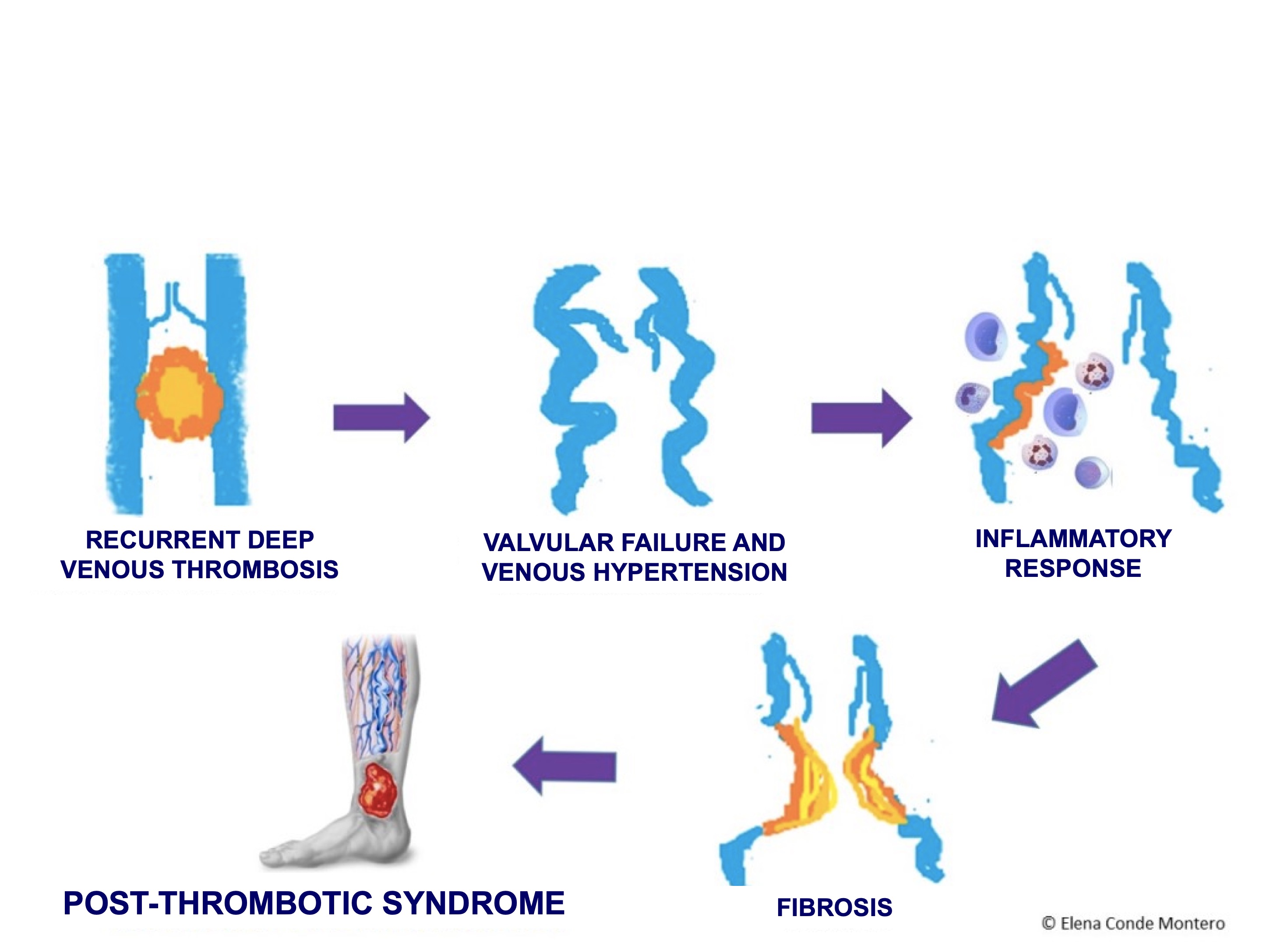
Many of the ulcers we may find in our daily clinical practice develop in the context of a post-thrombotic syndrome. It is important to know this entity, which has an important impact on the quality of life of patients and on health costs.

What is the post-thrombotic syndrome?

It is a long-term complication of deep vein thrombosis (DVT). Repeated episodes of DVT are a clearly established risk factor. It is the result of venous hypertension that develops due to valvular incompetence, reflux and/or venous obstruction, with an associated alteration in microcirculation. After the thrombotic episode, there is a weakening of the venous wall, with incomplete valve closure, reflux and secondary venous hypertension. Endothelial distension occurs, with increased wall permeability and chemotherapeutic effect on inflammatory cells, predominantly macrophages and leukocytes. The inflammatory process triggers a perivenous fibrosis and development of trophic alterations in the affected segment (See figure).

Its incidence is difficult to establish due to differences in follow-up time, patient selection and definition of post-thrombotic syndrome in the different studies. It is estimated that, one year after the occurrence of a deep vein thrombosis, up to 50% of patients will suffer post-thrombotic manifestations (Gabriel, 2003).

What are its clinical manifestations?

Among the signs that may be present, we find:

  • Edema
  • Tortuous veins
  • Skin alterations due to stasis. Ulceration would be the extreme degree, but hyperpigmentation, eczema and hypodermitis are very frequent findings. The development of ulcers may be triggered by trauma or may appear spontaneously. They are characteristically chronic with a high rate of recurrence.

The most common symptoms are pain, itching and a feeling of heaviness. (Torres, 1995).

How is it diagnosed?

Diagnosis is made through clinical and complementary macrocirculatory tests (color Doppler ultrasound, phlebography and magnetic angioresonance) and microcirculatory ones (Doppler laser flowmeters, transcutaneous measurement of partial pressure of O2 and CO2 and impedance plethysmography) (Gabriel, 2003). 

How is it treated?

Medical treatment:

In those patients  without surgical indication (most of them), local treatment of the ulcer must be associated with other strategies that can promote healing. The most widely accepted and used is compression therapy.

As we commented before, the role of microcirculation alterations in this syndrome is as important as changes in macrocirculation. Therefore, different drugs have been used that act at that level, favoring an adequate blood flow.

My experience is satisfactory with the use of either pentoxifylline and sulodexide, associated with compressive therapy.

The usefulness of both drugs in the treatment of chronic venous ulcers is supported by the results of different studies. Pentoxifylline has haemorrheological properties (increases the malleability of the red blood cell membrane) and inhibits the adhesion of platelets and leukocytes to the endothelium (Jull, 2012). Sulodexide is an orally administered heparinoid with antithrombotic and fibrinolytic action (Andreozzi, 2012).


Interventional treatment:

Depending on the hemodynamic findings, there are cases that may benefit from an invasive intervention. Correction of reflux points and improvement of venous return pathways may promote healing of skin ulcers.

Depending on the location and extent of the involvement, a variety of techniques can be used. When deep venous reflux is mild, saphenous vein ligation can eradicate the reflux in the femoral vein and achieve skin ulcer healing. However, if the reflux is severe, direct surgery of the deep system will be required. Valve repair achieves high healing rates, in some studies similar to those obtained in primary valve repair (60% absence of recurrence at 5 years) (Raju, 1996). However, the potential thromboembolic complications of this technique limit its use.

Preventive measures:

Given the absence of effective treatments for manifestations of post-thrombotic syndrome, prevention is essential.

Elastic compression stockings are the prevention measure considered most effective at present, with a 50% reduction in post-thrombotic syndrome. These data are the conclusion of 2 clinical trials with the use of compression therapy for 24 months (Brandes, 1997; Prandoni, 2004).  A recent clinical trial suggests that there is no benefit from wearing compression stockings (Kahn, 2014). However, these findings may be due to the high rate of non-compliance due to the discomfort of wearing compression stockings. The optimal indication and duration of compression therapy remains to be defined (Ten Cate-Hoek, 2010). 


References

Andreozzi GM. Am J Cardiovasc Drugs. 2012 Apr 1;12(2):73-81.

Brandjes DP, Buller HR, Heijboer H, et al. Randomised trial of effect of compression stockings in patients with symptomatic proximal-vein thrombosis. Lancet 1997;349:759–62.

Gabriel Botella F., Labiós Gómez M., Portolés Reparaz O., Cabanes Vila J. Nuevos avances en el conocimiento del síndrome postrombótico. An. Med. Interna 2003; 20(9): 45-54.

Kahn SR, Shapiro S, Wells PS, et al. Compression stockings to prevent post-thrombotic syndrome: a randomised placebo-controlled trial. Lancet 2014;383:880–8.

Jull AB, Arroll B, Parag V, Waters J. Pentoxifylline for treating venous leg ulcers. Cochrane Database Syst Rev. 2012 Dec 12;12:CD001733.

Prandoni P, Lensing AW, Prins MH, et al. Below-knee elastic compression stockings to prevent the post-thrombotic syndrome: a randomized, controlled trial. Ann Intern Med 2004;141: 249–56.

Raju S, Fredericks RK, Neglén PN, Bass JD. Durability of venous valve reconstruction techniques for “primary” and postthrombotic reflux, J Vasc Surg. 1996. 23: 357–366.

Ten Cate-Hoek AJ, Ten Cate H, Tordoir J, et al. Individually tailored duration of elastic compression therapy in relation to incidence of the postthrombotic syndrome. J Vasc Surg 2010;52:132–8.

Torres R. Clínica y diagnóstico diferencial de la trombosis venosa profunda y del síndrome postrombótico. In: Güell J, Rosendo A, editors. Enfermedad Tromboembólica venosa ( E.T.E.V.). Síndrome Postrombótico. Barcelona: EDIKA MED; 1995, p. 21-46.