Home Blog

The science of wound healing, my book

For all those who love reading books on paper, in my new book “The science of wound healing” you will find the most outstanding contents of my blog, with lots of iconography, clinical cases and therapeutic pearls.

The science of wound healing can be found on Amazon.com in the following links:

US: https://www.amazon.com/dp/B0CPYWJ3PX

UK: https://www.amazon.co.uk/dp/B0CPYWJ3PX

Best wishes for the New Year! Thank you for your trust and love:)

Leg ulcers due to heart failure?

Heart failure is the most frequent reason for hospitalization and readmission in the elderly. Although we do not usually think of it as a potential trigger for leg wounds, the reality is that it is a frequent cause of the appearance or worsening of leg ulcers. I am sure that reading this will have brought to your mind a case from the practice 😉

Why may heart failure trigger leg wounds?

Peripheral edema is a typical sign of congestive heart failure and, in episodes of decompensation, this edema can develop rapidly.

It is a bilateral edema, which often appears in combination with obesity, phlebolymphedema or hypoalbuminemia.

This edema may be related to left and/or right heart failure, to the autoregulation mechanisms of the cardiovascular system, as well as to concomitant fluid retention and/or to the medication used to treat it. In patients with right heart failure, blood stasis occurs, with swelling and accumulation of peripheral fluid. In left-sided heart failure, the increase in cardiac output, in addition to the possible influence on the deterioration of right-sided cardiac function, also activates compensatory mechanisms (nervous and hormonal) that result in peripheral edema related to the action of the renin-angiotensin system and sodium and water retention.

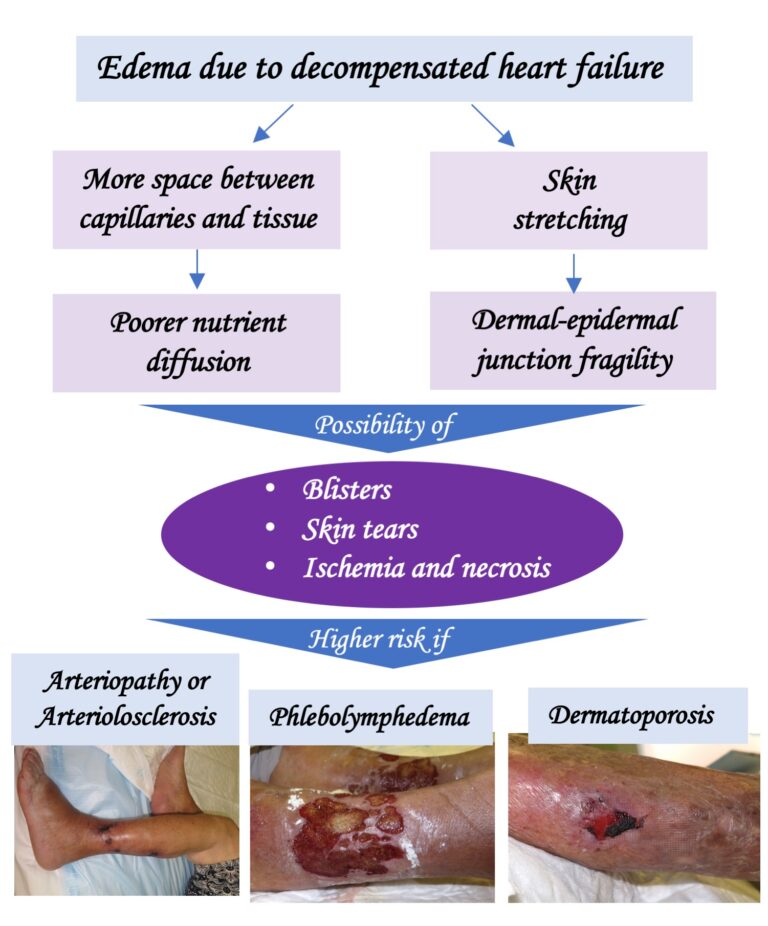
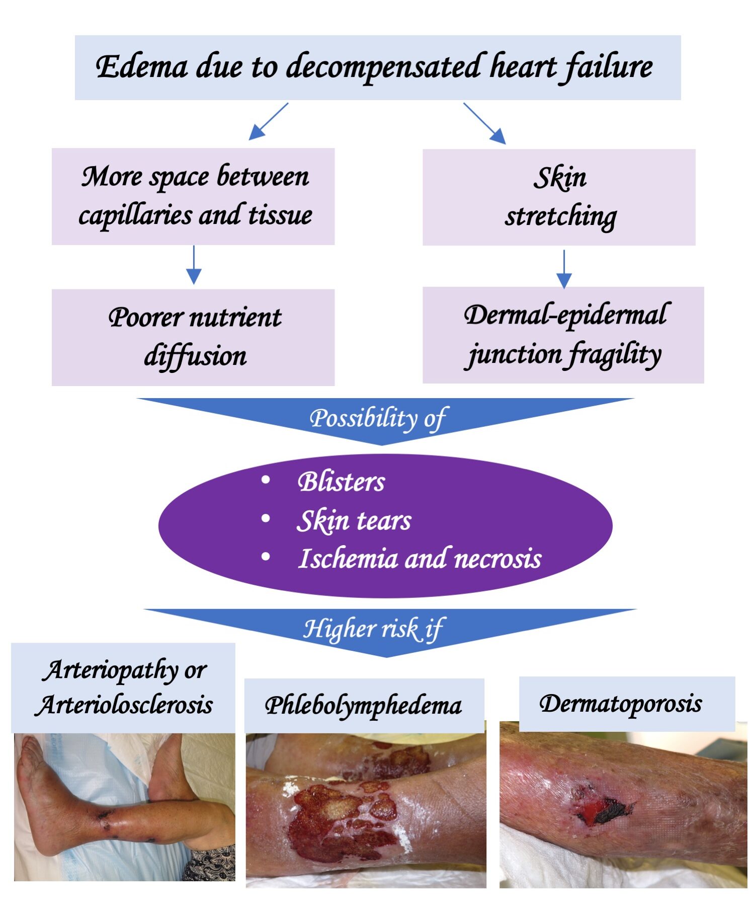
Whenever there is edema in the legs, regardless of its cause, the space between the capillaries and the tissue increases. This leads to poorer diffusion of nutrients and, as a consequence, decreased skin perfusion. Therefore, especially in patients with arterio/arteriolosclerosis or phlebolymphedema, in whom perfusion is already deficient at baseline, perfusion may become insufficient and cutaneous ischemia and necrosis may develop. In fact, it is not uncommon for Martorell ulcers (ulcers due to subcutaneous ischemic arteriolosclerosis) or arterial ulcers to develop after an episode of decompensated heart failure. On the other hand, heart failure is considered to be a risk factor for venous ulceration.2

In addition, fluid accumulation in the legs involves skin stretching and the possibility of detachment at the dermoepidermal junction, with the appearance of fissures, erosions and/or blisters. The condition will be more severe in people with dermatoporosis (see post: “Wounds secondary to dermatoporosis or chronic cutaneous insufficiency”), in whom skin tears will easily appear.

Bearing in mind that heart failure is a syndrome typical of the elderly, it is not uncommon for the same person to present together a certain degree of arterio/arteriolopathy, chronic venous and lymphatic insufficiency and dermatoporosis, which increases the risk of ulcers with a torpid evolution.

How should these wounds be managed?

The essential thing is to correct the cause of the edema, that is, to control heart failure, with diuretics and other drugs, for which the role of the internist is fundamental (I feel very fortunate to work surrounded by internists).

In order to be a pillar in the treatment of these ulcers, it is important that the diagnosis includes heart failure as a precipitating factor. For example: “Martorell ulcer in the context of an episode of decompensated heart failure”.

After correction of cardiac decompensation, compression therapy adapted to the needs and tolerance of each individual (provided there is no severe peripheral artery disease) will be of beneficial,1,3 starting with low pressures and devices with low resting pressure (short-stretch bandages, multicomponent systems, self-adjusting wraps with Velcro-type closure). Of course, anti-edema measures (rest with legs elevated) and muscle activation with dorsiflexion and plantar flexion exercises are also fundamental, even in patients with reduced mobility (See post: “What exercises can I do at home against phlebolymphedema?”) These measures are also important in a preventive manner, associated with adequate follow-up and control of heart failure. Taking into account that these people are usually elderly, with phlebolymphedema and/or skin fragility, we will preferably recommend skin protection socks and self-adjusting compression wraps (“Why are adjustable compression wrap devices so popular?”).

As discussed in the post “New recommendations about compression therapy“, it is recommended not to apply compression in severe cases of heart failure (NYHA IV = inability to perform any physical activity without discomfort, with symptoms even at rest). In less severe cases, the progressive increase in compression produces only very short phases of increased cardiac load and can facilitate a substantial reduction in peripheral edema.1

Depending on the comorbidities and underlying etiology, we may consider strategies that accelerate healing, such as negative pressure therapy or punch grafting.

A final thought: “How many people are suffering from leg wounds with poor evolution due to inadequate control of heart failure?

References:

  1. Urbanek, T., Juśko, M., & Kuczmik, W. (2020). Compression therapy for leg oedema in patients with heart failure. ESC Heart Failure, 7, 2012 – 2020.
  2. Augey F, Pinet A, Renaudier P. Insuffisance cardiaque et ulcère de stase : une association significative (étude prospective de 100 cas) [Heart failure and stasis ulcer: A significant association (prospective study of 100 cases)]. Ann Dermatol Venereol. 2010 May;137(5):353-8.
  3. Saucedo D, Evans N, Lim CS. Initiating compression therapy for those living with heart failure. Br J Community Nurs. 2021 Sep 1;26(Sup9):S12-S17.


6 reasons why a venous ulcer does not heal despite compression therapy

Compression therapy is the mainstay of treatment for venous ulcers. In fact, it is very accurate to say that, “when faced with a venous ulcer, without compression there is no option for healing“.

 However, there are times when, despite adequate compressive therapy, the evolution is not positive, since we do not manage to control the edema and/or the ulcer does not decrease in size. In this post we will analyze the situations that may be negatively affecting the wound and how to address them.

1- The pressure exerted is not sufficient or is not adapted to the needs of the person and the wound.

This occurs mainly in the following cases:

The morphology of the leg is very altered by lipodermatosclerosis and obesity, so there is not an adequate pressure gradient between ankle and calf. In this situation we must pad the leg to homogenize the perimeters (See post: “Band and bandage: not the same thing“).

The location of the ulcer is retromalleolar and the bandage or stocking makes a “tent” effect in this location. Therefore we should use a pad for local pressure or bandage strips specifically for local pressure. Here is a recently published article on the experience with these strips in the retromalleolar region.

The frequency of bandage change is not sufficient to adapt to the pressure drop secondary to edema reduction. This tends to happen when we use short stretch bandages in people with high mobility and edema, because of their great effectiveness in the rapid decrease of edema. The solution is to increase the frequency of bandage change or to use multicomponent systems, with a cohesive elastic bandage, which favors the maintenance of the bandage in place.

2- Venous reflux control is not achieved.

In these cases, the Doppler ultrasound test will identify incompetent veins that require treatment, either surgical or endovenous. Endovenous treatments, such as sclerotherapy, are minimally invasive and their objective is to eliminate the reflux and, therefore, the cause of venous hypertension.

The echo-Doppler test will identify incompetent veins that require treatment. The benefit of sclerotherapy is not limited to ulcers with isolated superficial venous insufficiency. Its use in ulcers with an arterial component or with associated deep venous system involvement can help control venous hypertension, with a decrease in edema and with a direct impact on healing (see post: “Sclerotherapy wins against recurrence of venous leg ulcers“).

3- Continued sitting with the legs in decline

When the person’s habit is to spend most of the day sitting with the legs down, compression therapy is not enough to control venous hypertension. In fact, the hydrostatic pressure in the legs during sitting is almost as high as during standing, as this article shows. Therefore, venous ulcer patients (and everyone else, as a generalized recommendation) should elevate their legs when sitting. Other lifestyle changes such as a healthy diet, physical exercise and weight loss will also have a positive impact.

In fact, exercise is essential to activate the plantar and calf pump, and thus promote venous and lymphatic circulation. As we commented in the post “Band and bandage: it is not the same thing”, the higher the stiffness index of the compression system used, the more effective it will be in resisting muscle contraction and achieving high pressure peaks that create brief and intermittent venous occlusions, similar to physiological valvular functioning. As muscle contraction is so important, muscle atrophy or limited mobility may decrease the effectiveness of compression bandages.

In case of phlebolymphedema and immobility (wheelchair), if there is also some degree of arteriopathy (ITB>0.5), the bandage with a high stiffness index with pressures lower than 30 mmHg (or the Velcro-type closure wraps) is a safe and effective option if dorsiflexion and plantar flexion exercises are performed. (See post: “Phlebolymphedema: a term that should be more used“).

4- The wound is “superchronic”

The chronicity of the wound, i.e. a long time of evolution, also has a negative influence on the response to treatment.

Why is this so?
Whereas in an acute wound the cells divide rapidly, in a long-standing ulcer, the fibroblasts of the wound bed and the keratinocytes at the edges become senescent cells, i.e. cells that, due to proinflammatory signals maintained over time, stop dividing.

In addition, slougyh tissue that is chronically maintained in the wound is associated with an increased risk of resistant biofilms) and chronic wound edges tend to thicken and lichenify, which hinders epithelialization.

On the other hand, other factors that hinder healing are phlebolymphedema, the woody consistency of the perilesional skin due to lipodermatosclerosis, hemosiderin deposits that produce ochre dermatitis (see post: “Iron, ochre dermatitis and venous ulcer”) and dystrophic calcifications that can appear secondary to chronic inflammation (see post “Why do calcifications occur in chronic skin ulcers?“).

  • So how can we help new tissue formation in these superchronic ulcers?

Sharp debridement and antimicrobial dressings will help fight biofilm.

In German-speaking countries, when lipodermatosclerosis is very intense, shave therapy is performed, which consists of excision of the ulcer and its inflammatory base in the hypodermis.

If there are dystrophic calcifications, their removal is essential to achieve wound closure.

Portable negative pressure therapy devices can stimulate granulation.

Skin grafts will promote epithelialization. Negative pressure therapy can facilitate graft attachment. Sometimes the procedure has to be repeated several times until complete epithelialization is achieved (See post: “Punch grafting and negative pressure therapy: a successful couple“).

5- Poor evolution is due to the occlusive effect of the dressings and bandage.

In cases where the problem is occlusion, the solution is to remove the dressings and bandages and leave the legs uncovered. To control edema in parallel, it is essential to rest with the legs elevated, ideally in bed with pillows under the legs.

This is not uncommon in wounds covered with punch grafts. In spite of adequate compression and rest with the legs elevated, sometimes in successive dressings during the first weeks we do not achieve complete epithelialization, with excessive exudate and a “congested” appearance of the grafts. Our experience is very good with the legs elevated and the application of zinc oxide drying solution once or twice a day on the grafted wound (See post: “In which situations would it be better to remove the bandage and leave the legs elevated and open air”).

6- When it is not a venous ulcer

The following statement is applicable to any wound: “If the response to a correct etiological treatment is not positive, you have to rethink the diagnosis of the wound”.

Look at the location, wound edges, wound bed tissue, perilesional skin… If you see atypical features that you would not expect to find in a venous ulcer, that person will benefit from dermatologic assessment and a skin biopsy… The differential diagnosis of venous ulcers is broad, and includes tumor ulcers (see post: “In front of a skin ulcer, when should we suspect a malignant ulcer?“).

In addition, it should be taken into account that, despite a predominantly venous ulcer, there may be some degree of associated arteriosclerosis (called mixed ulcer) or arteriolosclerosis (see post: “Little is said about arteriolosclerotic ulcers“).

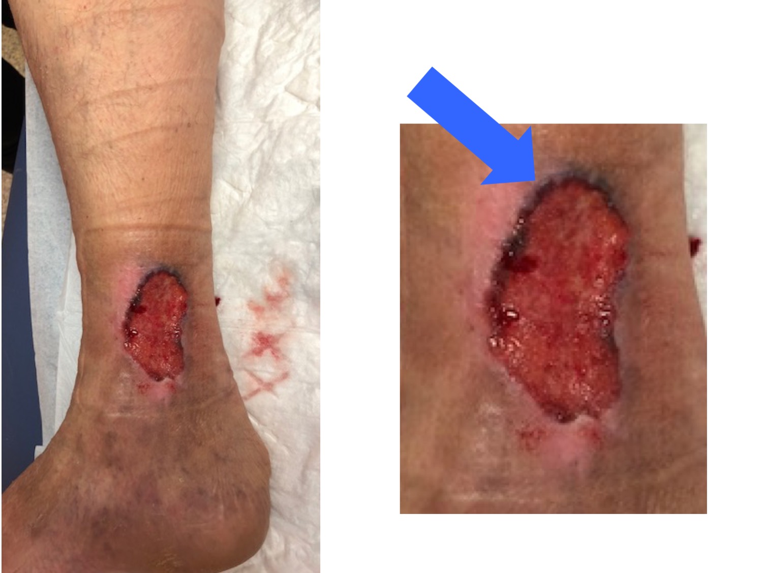
This is an example of a wound treated for many years as a venous ulcer, with compression therapy, without improvement, because it was a basal cell carcinoma (note that hyperpigmented raised border;).
I hope this post has provided you with some tips to continue fighting the damn venous ulcer;)) I take this opportunity to encourage you to read this consensus document that we published in 2019 on the management of hard-to-heal wounds:):)


Why punch graft attachment has not been achieved?

This is a question that you will have asked more than once all of you who are performing this technique:)

As we have commented in other posts, punch grafts, even if they do not adhere to the wound bed, release growth factors and cells that, in addition to promoting healing, also reduce pain. (See post: “Punch grafting as a painkiller for painful ulcers“) That is why its list of “unofficial” indications is so extensive and can include arterial ulcers that are not candidates for revascularization, when the patient does not want amputation.

So, taking into account the wide profile of ulcers that we can treat with punch grafting, we must be open-minded when evaluating success or failure. What we should never graft are infected wounds or ulcers in which we have not optimized the etiological treatment.

We speak of attachment when, at the first dressing change from the 4th day of the procedure (ideally, one week later), the grafts are adhered to the bed and present a pink-bluish coloration, a sign that neoangiogenesis (formation of new vessels that nourish the dermis and epidermis) has occurred.

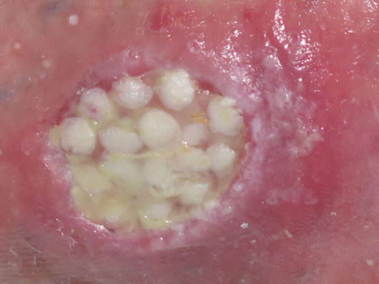
Well, this ideal attachment in the first week occurs when we graft a bed in optimal conditions … That is, rarely … Not to say almost never … 😉 Therefore, it is normal in clinical practice that, especially when grafting wounds with some slough (usually it is not possible to achieve an optimal bed), we find grafts with yellowish or whitish coloration that remain in the bed by imbibition. In this situation we will not do any type of cleaning on the bed and we will make a new cure a week later (if the exudate allows it). In the following dressing changes some grafts, or even all of them, may become detached 🙂


I would like to underline that in a study we conducted on venous ulcers, compressive therapy and punch grafts, ulcers with a percentage of sloughy tissue in their bed greater than 50% showed no less tendency to complete epithelialization:)

– And what do I do if from the first dressings there is no data of attachment and many of the grafts remain in the dressing?

The first thing you should do is to reposition in the wound the grafts that have remained in the dressing (See post: “Repositioning and not removing the scabs: two unwritten rules of punch grafting“). Speaking of dressing… There are no comparative studies on the usefulness of different dressings in the grafted area.

  • Secondly, in lower limb wounds, you should ask the patient and family members if they have been on absolute bed rest. Absolute bed rest with the legs elevated, at least the first 4 days (although we always recommend a minimum of one week), is essential to promote graft attachment in lower limb wounds. In fact, this is the main cause of graft failure in our practice, which forces us to admit those patients who cannot perform adequate rest at home.
  • Thirdly, reconsider whether the problem may have been inadequate bed preparation prior to coverage. Before the next coverage, make sure to minimize the presence of biofilm.
  • In case we find complicated locations, such as the Achilles tendon or the malleolus, or beds with a lot of slough that we cannot remove or very undermined, we have to think that what may have failed is insufficient contact and pressure of the grafts with the wound, so the next coverage with grafts would be performed with ambulatory negative pressure therapy (See post: “Punch grafting and negative pressure therapy: a successful couple“).

  • If you have grafted an ulcer in the spectrum of arteriolosclerosis, for example, a Martorell ulcer, early, with active necrosis and abundant slough, think that your objective was not to achieve attachment with those first grafts, but a reduction of pain, control of necrosis and improvement of the bed for the following coverings. Sequential grafts every 3 weeks or so will help us to achieve complete epithelialization in these cases (see post: “How to close large ulcers after minor trauma in the elderly“).
  • Finally, rethink the diagnosis of ulcers that, despite having been grafted in near-optimal conditions and with adequate etiological treatment, do not respond.

– And what has gone wrong when punch grafts take hold but in the following weeks are lost?

This usually happens in venous ulcers of long evolution. Resting with the legs elevated controls venous hypertension, but this is lost when the person begins to lead a normal life because, in spite of using compressive therapy, he does not take anti-edema measures, such as having the legs elevated during rest. A sign of venous congestion associated with venous hypertension is the maroon-purple color of these grafts prior to their detachment. It is essential to insist on the importance of anti-edema measures in people with venous ulcers!

– And when the problem is that, despite adequate compression and rest, in the successive dressing changes during the first weeks we do not get a complete epithelialization, with excessive exudate?

As I commented in the post “In which situations would it be better to remove the bandage and leave the legs elevated and open air?“, the problem in this case is occlusion. Therefore, the solution is to remove the dressings and bandages and leave the legs open air. To control edema in parallel, it is essential to rest with the legs elevated. Our experience is very good with the application of zinc oxide drying solution once or twice a day. To perform this strategy we usually need a hospital admission.


Have you already performed punch grafting? What is your experience?

Why does the classification of pressure ulcers fall short?

Because it does not include information that is very relevant in the evaluation of these wounds…

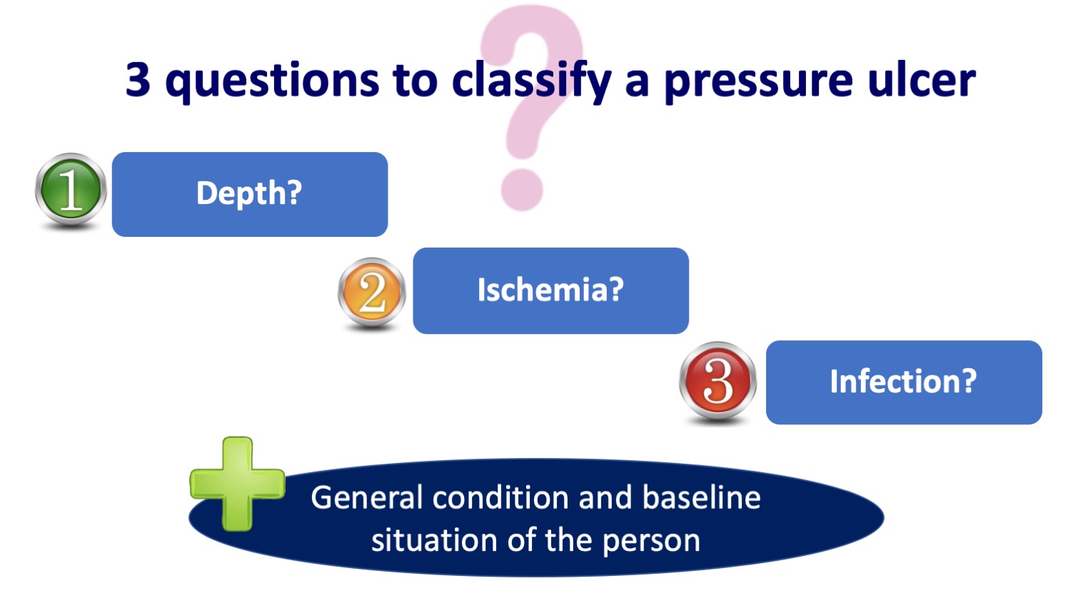
As a reminder, the most widespread and widely used classification of pressure ulcers is the NPUAP/EPUAP (National/European Pressure Ulcer Advisory Panel) system, which only includes the depth of the wound.

The problem is that talking only about the depth does not give us enough information to get an idea of the prognosis and decide the best treatment in each case… In fact, many times, the main conditioner of the evolution is ischemia or osteomyelitis, which are not recorded in this classification (See post “Do not debride dry eschars on heels“). And, of course, the general condition and baseline situation of the person.

Although I had already reflected many times on the limitations of this classification, I have been encouraged to write on the subject after reading two articles that have just been published by the working group of my friend and great expert in wounds, José Contreras, which you will find at the end of this entry. These authors propose a new classification of pressure ulcers including, in addition to depth, ischemia and infection. This classification is called DIP (Depth, Infection, Perfusion) and is inspired by the University of Texas diabetic foot lesion classification scale.

What are the most relevant practical aspects of this new DIP classification proposal?

  • Grade 0 represents a pre- or postulcerous lesion that includes healed ulcers, scars and non-blanching erythema. Although this grade does not describe a true ulcer, it helps identify sites at high risk for ulceration or re-ulceration.
  • Wounds with bone or joint involvement are included in a specific category (grade III), as they have a worse prognosis.
  • Grade III ulcers have a high risk of osteomyelitis, so this should be ruled out if the evolution is not favorable.
  • It is mandatory to palpate pulses in any person with a heel pressure ulcer.
  • The presence of ischemia, regardless of the depth of the ulcer, will require an assessment by vascular surgery.
  • In case of ischemia and absence of infection, debridement should be avoided and the wound kept dry if there is no possibility of revascularization. On the contrary, if there is ischemia and signs of infection, it is essential to remove the non-viable tissue to control the infection.
    Finally, I would like to stress that, although the characteristics of the ulcer will establish the treatment to be followed and are a prognostic marker, the mobility, mental state and general condition of the person are a fundamental pillar in decision making. In people at the end of life, the most important thing is to seek wellbeing, spacing the cures as much as possible without being invasive. In fact, this strategy of touching wounds as little as possible is the strategy of choice in Kennedy’s so-called terminal ulcers. Although their exact etiology is unknown, these rapidly progressive multiple pressure ulcers could be framed as multi-organ failure.


Have you heard about the “sitter´s sign?”

Most likely you don’t know the name of the sign, but if you work with older people, you’ve probably seen the lesions it describes on more than one occasion 😉 In fact, they are a frequent reason for consultation.

The “sitter sign” refers to the rough, thickened skin that older people often develop near the intergluteal cleft, associated with immobility and continued sitting. It is also known by other more complicated names, such as gluteal senile dermatosis or hyperkeratotic lichenified skin lesion of the gluteal region.1 The latter name, although more cumbersome, actually describes the disorder well:). Gluteal senile dermatosis was first described in Japan in 1979 and, since then, the few publications on this pathology are predominantly cases and case series from Asian countries.2,3 However, despite the lack of published studies in our setting, it is also very prevalent in Western countries.

What are the triggers?

The fundamental causes appear to be advanced age (thinning and loss of skin elasticity), prolonged sitting (with consequent pressure and friction) and a low body mass index.2,3 Affected individuals spend most of their time sitting. It would be interesting to study the prevalence of these injuries in young people who require continuous wheelchair use.

Although the first published case series show a striking predominance of the male sex, it is a pathology that is equally prevalent in women.2,3

How does it present clinically?

Typically, hyperkeratotic, scaly, ill-defined, brownish-red to grayish, poorly defined plaques slowly develop in the intergluteal cleft and on both buttocks. This characteristic distribution pattern of lesions is reminiscent of the “three vertices of a triangle”.2,3 In addition to darker, non-inflammatory plaques, inflammatory plaques with a psoriasiform appearance may be observed.

The most frequent, but not exclusive, location of plaques on the buttocks is in the area of the ischial tuberosities and lesions of the gluteal cleft do not necessarily coincide with the coccygeal apex. However, there are incomplete forms, as some patients have lesions only in the buttocks and not in the intergluteal cleft, or only in the intergluteal cleft, and sometimes only one buttock is affected. The appearance of hyperkeratotic ridges perpendicular to the buttocks has been described as typical.2,3

Erosions and ulcers of different depths may also appear.

They are usually asymptomatic lesions, but may cause discomfort when sitting, especially on unpadded surfaces. The discomfort includes itching, pain and foreign body sensation. Considering that in most cases they are asymptomatic and therefore do not cause consultation, it may be a much more prevalent pathology than is generally thought.

How is it diagnosed?

Diagnosis is clinical. We will only perform a biopsy to rule out other pathologies if we have doubts. In its differential diagnosis we will include irritative intertrigo, tinea, candidiasis, inverse psoriasis, eczema, lichen simplex chronicus and anosacral amyloidosis.2,4

If we biopsy, in most cases the histopathologic changes will be mild and characteristic, although not specific. Typical findings include orthokeratotic hyperkeratosis, epidermal hyperplasia and vascular dilatation in the papillary dermis with little lymphohistiocytic infiltration. In more severe cases, additional changes appear: edema of the papillary dermis, dilatation and proliferation of small vessels down to the reticular dermis and a dense lymphohistiocytic infiltrate, as well as erosions or ulcers.4

These findings indicate a type of abnormal keratinization resulting from chronic friction. Thus, the wounds that appear in senile gluteal dermatosis occur from the epidermis that is thickened, which differentiates them from pressure injuries, in which epidermal damage is secondary to necrosis due to ischemia and cellular stretching.

How is it treated?

Senile gluteal dermatosis often responds poorly to topical corticosteroid treatment. Topical corticosteroid may relieve itching but does not usually improve hyperkeratotic skin lesions. On the other hand, keratolytic agents, such as salicylic acid or urea, also do not seem to give satisfactory results. The benefit of topical retinoic acid has been described, as it may help to control abnormal epithelial differentiation and desquamation. The use of emollients to prevent skin dryness is also recommended, in addition to frictionless washing and drying of the area. When wounds have already appeared, my experience with zinc oxide barrier products is good.

However, the most effective therapeutic measure is patient and family education on the importance of implementing lifestyle changes that include avoidance of prolonged periods of sitting. In addition, pressure relieving devices during sitting (in severe cases ideally static or dynamic air cushions) may be helpful.2,3

References:

  1. Dahl, M.V. (2014). Sitter’s Sign. NEJM Journal Watch, 2014.
  2. Moon SH, Kang BK, Jeong KH, Shin MK, Lee MH. Analysis of clinical features and lifestyle in Korean senile gluteal dermatosis patients. Int J Dermatol. 2016 May;55(5):553-7.
  3. Liu HN, Wang WJ, Chen CC, Lee DD, Chang YT. Senile gluteal dermatosis: a clinical study of 137 cases. Int J Dermatol. 2014 Jan;53(1):51-5.
  4. Liu HN, Wang WJ, Chen CC, Lee DD, Chang YT. Senile gluteal dermatosis – a clinicopathologic study of 12 cases and its distinction from anosacral amyloidosis. J Eur Acad Dermatol Venereol. 2012 Feb;26(2):258-60.

When sharp debridement “angers” the ulcer

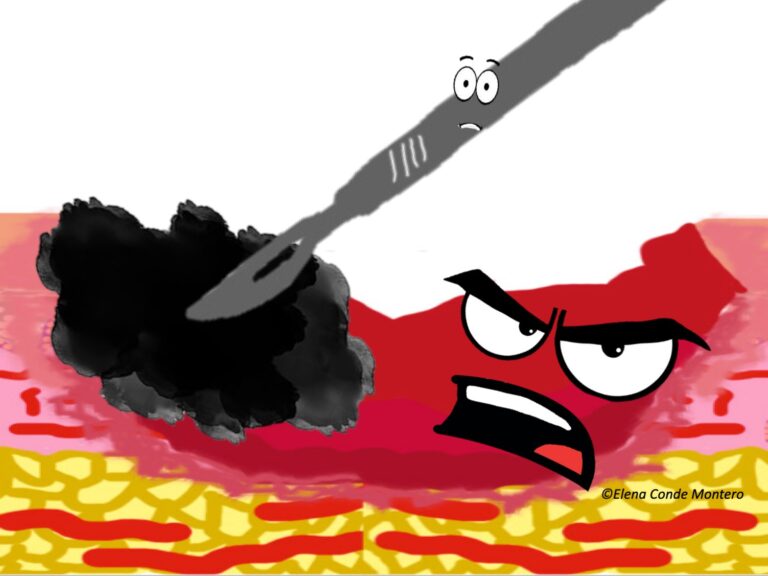
Sharp or surgical debridement of an ulcer involves the use of a scalpel blade to remove necrotic or sloughy tissue. Unlike other types of debridement, such as autolytic or enzymatic debridement, it has the advantage of speed in removing nonviable tissue, sometimes in a single session. But it also has its disadvantages, such as possible bleeding or pain, and certain contraindications.

The three best known contraindications are: severe peripheral artery disease until it has been revascularized, dry necrotic plaques on heels and active pyoderma gangrenosum. Let’s look at these last two in more detail….

Why should we not remove dry necrotic plaques on the heels?

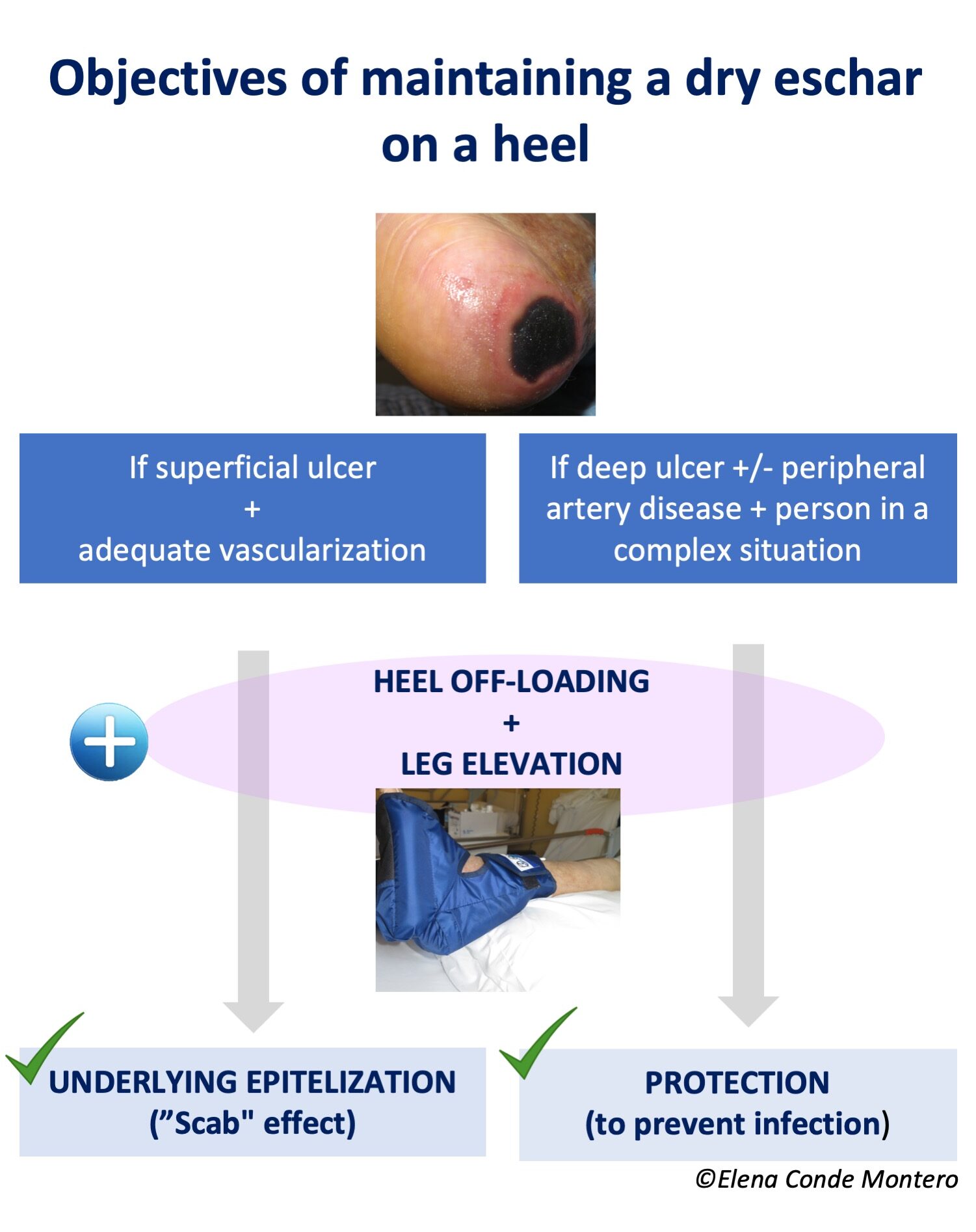
Because being dry, adherent to the bed, without fluctuation or other signs of infection, they have a protective function. Keeping that protective plaque dry can prevent bacterial proliferation and have a “scab effect”. In addition, it avoids the pain of sharp debridement and the possibility of leaving exposed deep tissues (calcaneus) with potential for infection.

If the ulcer is superficial, not very extensive and the vascularization is adequate, this scab could promote the underlying epithelialization. On the contrary, if the ulcer is deep and extensive, there is associated peripheral arterial disease and/or the patient’s complex situation does not allow another attitude that favors healing, the objective will be exclusively to maintain the “protective scab” to avoid superinfection (palliative protection or prior to revascularization or amputation). See post: “Do not debride dry schars on heels“.

What is the phenomenon of pyoderma gangrenosum patergia?

Pyoderma gangrenosum is characterized by uncontrolled activity of the immune system, predominantly neutrophils, with production of necrosis and skin ulcers. It may appear spontaneously or be triggered by minor trauma or surgery. This inflammatory hyperreactivity is called patergia phenomenon and can appear after sharp debridement (See post: “Pyoderma gangrenosum: a diagnostic and therapeutic challenge“). In fact, debridement can rapidly increase the extent and depth of the ulcer. To avoid this, we will only perform this type of debridement when immunosuppressive treatment has adequately controlled the inflammatory activity and there are no signs of activity (See post: “How to promote healing in ulcers secondary to pyoderma gangrenosum?”).

The three contraindications we have just seen are the most agreed upon. However, there are other situations in which sharp debridement can worsen wound evolution, such as Martorell ulcer or ulcers secondary to vasculitis.

Why can a Martorell ulcer be worsened by sharp debridement?

Extensive sharp debridement can behave like trauma, with a first phase in which vasoactive substances are released to produce vasoconstriction to limit tissue damage, bleeding and facilitate clot formation. To this end, the arterioles limit the flow by contracting. Following this vasoconstriction, there is significant vasodilatation with increased permeability of the arteriolar wall, so that cells (neutrophils, macrophages) and inflammatory molecules are provided to the wound bed.

Patients with Martorell ulcer present subcutaneous arteriolosclerosis, i.e., less reactivity of the arteriolar wall, calcifications and decrease in caliber, this vasoconstriction may imply a compromise of tissue irrigation prolonged in time, with consequent ischemia. Subsequent vasodilatation with release of inflammatory mediators increases necrosis (cell death), and here the vicious circle of necrosis-inflammation begins. Necrosis secondary to this inflammation would maintain the pro-inflammatory mediators with consequent extension of the wound.

This occurs not only in Martorell ulcer, but in the whole spectrum of arteriolosclerosis ulcers (See post: “Little is said about arteriolosclerosis ulcers“).

BEWARE! This possible worsening with sharp debridement, in addition to the frequent finding of an abundant inflammatory infiltrate in superficial biopsies, may lead to misdiagnosing of Martorell ulcer with pyoderma gangrenosum.

Taking this into account, although sharp or surgical debridement is considered the first choice prior to graft coverage, in our practice we try to be as conservative as possible, with minimally invasive debridement and early and sequential punch grafting (see post: “Interest of early punch grafting in Martorell hypertensive ischemic ulcer“).

What about vasculitis?

It is not uncommon to see increased necrosis after sharp debridement in the acute phase of vasculitis in which, in addition to inflammation, there is vascular occlusion. This worsening could be framed within the concept of “vicious circle necrosis-inflammation” that we have just explained. Therefore, as in pyoderma gangrenosum, in ulcers due to vasculitis it is recommended to wait until immunological activity is adequately controlled by immunosuppressive treatment. As in the case of pyoderma gangrenosum, the control data of the inflammatory activity are not always easy to identify…

I would like to end with a therapeutic pearl that can facilitate sharp debridement of necrotic plaques. It is a drying strategy to make the subsequent removal of the plaques with the scalpel less traumatic and painful 😉

The idea to write this post came to me in a session on atypical wounds at the World Union of Wound Healing Societies Congress just held in Abu Dhabi:) I am happy to be back at congresses. There are many ways to share knowledge, but in person will always be more enriching. Life is about meeting people;)

Zinc oxide and alginate: a perfect combinatio

I have just returned from the joint EWMA& Journées Cicatrisations 2022 congress held in Paris. I thought I would dedicate a post to the new technologies that have been presented and are already available on the market. I would have to talk about cold plasma, phototherapy, electrical stimulation, oxygen therapy, new skin substitutes, sensors for the early detection of pressure ulcers, thigh compression device as an adjuvant to leg compression…. However, I have decided to leave these innovations on hold until such time as I have some experience with some of them and can share my experiences with you:)

As you know, I love the necessary innovation, but I also believe that it is essential to rescue traditional products and practices that have proven to be efficient. It is not because a therapeutic strategy is “old” that it will be worse than a new one. Although I have already dedicated separate blog posts to them (“Why do we use so many alginate fibre sheets in our wound clinic?”, “Why do we use topical zinc on wounds and perilesional skin?”), alginate and zinc oxide deserve a post together because we combine them so much in the practice:)

Why is the combination of zinc oxide and alginate of interest?

Alginate is a polysaccharide formed by mannuronic acid and guluronic acid units. It has demonstrated biocompatibility, non-toxicity, non-immunogenicity, biodegradability, hemostatic activity and can simply gel. When dried it can simulate a “scab effect” that will promote underlying epithelialization.

Although the exact mechanism of zinc in wound healing is unknown, as it is essential for the activity of many enzymes, it appears to be involved in cell replication and migration, protein synthesis, cell repair and autolytic debridement. It has also been associated with an anti-inflammatory and antimicrobial effect.

Zinc oxide allows a sustained release of zinc while avoiding cytotoxic levels. It seems to simulate the action of certain growth factors, as well as increasing their release.

Bandages combining alginate and zinc oxide have been developed, with improved healing and reduction of bacterial flora (see article).

But this combination can also be done in the office, using products with zinc oxide and calcium alginate sheets. Although the best known pharmaceutical form of zinc oxide is in a paste, as a barrier product, in exudative lesions we will use an aqueous base, due to its drying action. Before applying zinc oxide lotion, it must be shaken, as it does not dissolve in water.

In what situations do we use this combination in our wound practice?

In all those cases in which we need to control inflammation, promote epithelialization, control exudate and promote a scab effect. That is to say, among its main uses are:

Acute, superficial, uncomplicated wounds. An example is the graft donor site, a type of wound that we produce every day in the office, or traumatic wounds, such as skin tears.

Moisture-associated lesions. These superficial erosions and ulcers can appear in folds (irritative intertrigo), areas of friction and pressure (sacral and gluteal region), or in the perilesional skin due to wound exudate. If there is a high inflammatory or eczematous component, we associate topical corticosteroid.

-Friction blisters, for example, secondary to shoe rubbing or slipping of a compressive bandage. In these cases, prior to covering with zinc oxide and alginate, we puncture the blister and empty its contents while maintaining its roof. Maintaining and repositioning the detached epidermis helps to promote epithelialization and reduce pain.

Grafted ulcers or ulcers in the epithelialization phase. This combination promotes complete eplithelialization and prevents maceration.

Second degree superficial burns. Taking into account that burns have an excessive inflammatory component (responsible for pain, delayed healing and pathological scarring), we add medium-high potency topical corticosteroid to the combination of zinc oxide and alginate. See post: “Does it make sense to apply topical corticosteroids to improve the healing of superficial burns?”)

How do we cover the alginate sheets?

This is a very important question, since we need a fixation that allows us to space the dressings as much as possible and that does not damage the perilesional skin.

In cases of lesions associated with moisture, the ideal is not to place any occlusive dressing over the alginate, i.e. nothing or only gauze.

In case of wounds in locations other than the leg, we will use a gauze with adhesive or a polyurethane film, which will be more atraumatic, especially in fragile skin. In the leg, the coverage will be with a bandage or compression stocking (See post “Compression is key to treating leg wounds”).

The more we space the dressings, the less we will interfere in the epithelialization process and the less risk of “tearing” of the neoformed epidermis during the removal of the dressing. This is especially important in skin tears in the elderly, where we could keep the dressing on without removing it for three weeks, until complete epithelialization.

Why do wounds itch?

How many times have you heard “if a wound itches it is because it is healing”? Well, this popular belief has some truth to it, since itching is normal in the healing process and usually appears in the later stages. However, as we will see below, it occurs in both acute and chronic wounds, and even scars, and is mainly associated with the inflammatory process (pruritoceptive pruritus) or nerve damage (neuropathic pruritus), not necessarily with a good wound evolution.

Itching  can be very bothersome and unresponsive to multiple treatments. This can have a great impact on people’s quality of life, especially when it is persistent in chronic wounds or scars.1-5

There are still many unknowns about the pathophysiology of wound pruritus, but in this post we will summarize the available evidence that helps us to better understand this very uncomfortable symptomatology.

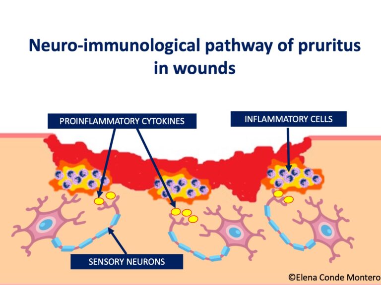
Proinflammatory cytokines released in any wound (acute or chronic) can activate sensory neurons and produce itching. This is termed the neuro-immunologic pathway of pruritus and multiple cells appear to be involved, including white blood cells, keratinocytes and endothelial cells.4 Pruritogenic molecules such as interleukin 31(IL-31)3 activate afferent sensory fibers in the skin and these neurons synapse with second-order neurons in the spinal cord. After communication with a complex of spinal interneurons, a signal is sent to the brain where the itch sensation is recognized. Instructions are then sent to the motor neurons responsible for scratching.

Microenvironmental factors also contribute to itching. These include dry scabs and perilesional deramatitis, as well as the presence of bacteria in the wound bed.

Histamine does not seem to be a clinically relevant contributor to wound-associated itching, so the interest of antihistamine use would be practically limited to its sedative effect.

A prospective study involving 200 chronic wounds of different etiologies (traumatic, pressure, neuropathic, venous, arterial, mixed and others) found that 28% of patients had wound-related pruritus. Lower extremity wounds were more likely to be itchy than upper body wounds and venous ulcers were significantly more itchy than other types of chronic wounds. Wound duration did not predict the occurrence of itching; however, pruritus was more prevalent in larger wounds, with edema, more granulation tissue or necrotic tissue.5

The persistence of pruritus after a burn is striking. The precise trigger for this phenomenon is unknown, but it seems that the neuropathic mechanism is the main one involved. In fact, these patients may report paresthesias (needle pricks). Neuropathic pruritus is due to increased sensitivity and excitability of damaged peripheral afferent fibers, which form neuromas. It may therefore respond to neuroleptic agents such as gabapentin and pregabalin.4

In pathologic scars following wounds with an intense inflammatory phase, i.e. hypertrophic and keloid scars, chronic pruritus is not uncommon. In keloids, abnormalities of afferent nerve fibers have also been reported, which could explain this itching.2

Proteomics and metabolomics studies will allow us to profile in more detail the mediators involved in wound itching and will help us understand why some wounds and scars itch and others do not. These data can then be used to develop targeted therapies for itch. Until then, wound itch treatment will remain poorly targeted.

References:

1. Lerner E. Why Do Wounds Itch? Wounds. 2018 Jan;30(1):1-3.

2. Parnell LKS. Itching for Knowledge About Wound and Scar Pruritus. Wounds. 2018 Jan;30(1):17-36.

3. Xu J, Zanvit P, Hu L, Tseng PY, Liu N, Wang F, Liu O, Zhang D, Jin W, Guo N, Han Y, Yin J, Cain A, Hoon MA, Wang S, Chen W. The Cytokine TGF-β Induces Interleukin-31 Expression from Dermal Dendritic Cells to Activate Sensory Neurons and Stimulate Wound Itching. Immunity. 2020 Aug 18;53(2):371-383.e5

4. Chung BY, Kim HB, Jung MJ, Kang SY, Kwak IS, Park CW, Kim HO. Post-Burn Pruritus. Int J Mol Sci. 2020 May 29;21(11):3880.

5. Paul J. Characteristics of chronic wounds that itch. Adv Skin Wound Care. 2013;26(7):320–332. 53. 


The color code of punch grafting

The technique of obtaining and applying grafts in the wound is very simple. However, to be successful with punch grafting, it is essential to know their “color code”;) In this post we are going to understand what the different colors that we can find in dressing changes after the procedure mean.

Graft coloration is associated with neoangiogenesis. As a reminder, neoangiogenesis is the formation of new vessels, which occurs in the first few days after the procedure. Until that time, the grafts do not have their own blood supply, so they must rely on the wound bed for nutrients. If you want to go deeper into the subject, you can read Braza ME, Fahrenkopf MP. Split-Thickness Skin Grafts. [Updated 2022 May 8]. In: StatPearls [Internet].”

Important notice before continuing: we can find all colors at once in a single wound! (In fact, it is a very frequent occurrence;).


– Yellowish-white:

This pale coloration of the grafts is due to ischemia because a vascular network has not yet been established between the cut vessels on the underside of the graft and the capillary beds in the wound bed. Grafts survive by imbibition, i.e., oxygen and nutrients from the wound bed reach the graft by passive diffusion. This coloration is normal during the first week after the process, usually turning pinkish or bluish in the following days as the graft revascularisation process is completed.

– Blue-purplish:

This is due to venous congestion of the grafts, which is a normal phase in the process of neoangiogenesis and, therefore, a sign of graft attachment. In the following weeks venous drainage of the new vessels occurs and the coloration becomes pinkish.

– Pink:

Indicates attachment, i.e. a vascular connection between the graft and the bed. It is common to find pink grafts intermingled with blue-purplish grafts and even pink and violet coloration in the same graft.

– Yellowish-brownish:

This is a sign that the grafts are maintained by imbibition, that is, by diffusion of nutrients from the bed but neoangiogenesis has not occurred. We usually find this coloration when we have grafted a wound in conditions that are not optimal. When we observe it after the first week after the procedure, and the pieces are not fixed in the bed (they are “dancing” in the bed), it indicates that these grafts will not take. Despite this lack of attachment, it is important to keep them in the wound bed, since they release growth factors and cells that promote healing and reduce pain. If we see a stagnation in the healing, from the third week, we can consider the removal of these grafts, which are not attached, and perform a new coverage with punch grafting.

It is also normal to find remains of exudate and scabs between the grafts, which have an epithelialization-promoting function, as well as a protective one. It is therefore essential not to remove this material, which can present different coloration, from yellowish-brownish to garnet-blackish.

After this explanation, you can now understand why the appearance of these grafts in the first dressing change, one week after the procedure, is normal;)