Home Blog Page 4

How to prevent and treat skin lesions cuased by PPEs

Personal Protective Equipment (PPE) is the main ally of health professionals in this COVID-19 crisis. That is why it is so important that they are always available and that their manufacture and maintenance comply with established safety regulations. Although their very name implies that they protect, we also have to protect ourselves from them… We especially have to prevent them from damaging our skin. I have been wanting to write about this for a long time, but I needed more experience, both personal and from other health professionals, as well as research into what has been published on the subject. Since I’ll be taking part in a webinar on skin lesions secondary to PPEs on April 30th, organized by the EWMA (to which, of course, you’re all invited), I think it’s high time to dedicate a post to it in the blog.

Knowing strategies for prevention and treatment of these injuries is essential, because when they appear they can be very limiting, with an impact on your quality of life and professional performance. Different scientific societies, hospitals and other health institutions are proposing and publishing recommendations and protocols for protection and treatment. Among others, I have found very interesting those of the British Society for Cutaneous Allergy & British Association of Dermatologists.

Before moving on to the injuries produced by protective goggles, masks and other devices that are part of this protective equipment, we have to deal with a problem that was already common among healthcare professionals, but which has increased exponentially in recent weeks: irritant hand dermatitis. This is due to excessive washing, the continuous use of hydro-alcoholic solutions and the prolonged use of several overlapping gloves, which alter the hydrolipidic layer of the skin and, therefore, cause the loss of the barrier function of the most superficial skin layer. People with chronic dermatitis, such as atopic dermatitis, are most affected. Although the degree and type of affectation is very variable (from dryness to fissures and erosions), and if there is any doubt or complication a dermatologist should evaluate you, there are a series of general measures that can be recommended:

  • It is recommended that you pat your hands dry, not rub them.
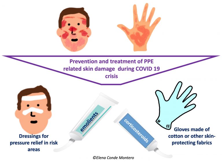
  • The application of unscented emollient products should be frequent, both in the hospital and at home. The use of paraffin products and coverage with cotton gloves or other skin protective fabrics is an interesting strategy during the hours of night rest to increase the penetration of the ointment and its moisturizing power.
  • The “best emollient” is the one with which one feels hydrated and comfortable.
  • If the application of moisturizing products is not enough and erythematous desquamative plaques, vesicles, lichenified lesions with fissures appear, it is important the dermatological evaluation since the use of topical corticosteroids of medium-high potency in cream or ointment during 1-2 weeks will help to control the outbreak. It should not be forgotten that, as in any case of irritant dermatitis, if its trigger cannot be eliminated (as it is in this case), the response to the treatment, or its duration, will be very variable.

We will now focus on the lesions produced specifically by the components of PPEs. A Chinese study on the prevalence of these injuries has just been published, in which 542 health professionals completed the surveys.1 The result was a prevalence of 97%. The most affected locations were the nasal bridge (most frequent, 83.1%), hands, cheeks and forehead. Dryness, tightness, scaling and erythema were the most common signs and symptoms. Another important result, which was to be expected, is that the risk of developing these lesions is greater in those professionals who are wearing the equipment (goggles, FFP2-FFP3 masks) for more hours. However, wearing face shield for more hours was not associated with more skin damage. This study also concludes that hand dermatitis is associated more with frequent washing than with prolonged use of gloves.1 The higher frequency of damage to the bridge of the nose is essentially explained by the pressure overlap of the goggles and mask.

Taking into account these findings described in the literature, which we also find in our daily practice in the hospital, here are some recommendations that may be useful:1-4

  • Promotion, as far as possible, of short shifts among professionals (e.g. every 2 hours) to temporarily relieve the pressure zones of the FFP3 mask and avoid excess humidity under the mask.
  • Use of fast-drying, residue-free barrier products that can prevent moisture damage (sweating) under the mask and other protective material.
  • To alleviate pressure and friction in risk areas, mainly the nasal bridge, but also the forehead and cheeks, we can use different atraumatic dressings to act as an interface. Hydrocolloids and silicone foams are the most common. However, we have to make sure that their use does not prevent an adequate adaptation and sealing of glasses and mask to the face. In addition, we must take into account that frequent application and removal of these dressings can also cause damage to the epidermis, the most superficial layer of the skin.
  • Taking into account that it is frequent the development of irritant contact dermatitis, with erythemato-descamative pruriginous lesions, it will be very important the facial moisturizing at home and, if necessary, the application of topical corticosteroid cream once a day during 3-4 days.
  • The daily use of facial moisturizer is essential to protect the skin barrier. However, it is recommended that it be done at least 30 minutes before the application of the mask and other equipment to ensure its fixation and avoid altering its composition.
  • Since it is frequent that those people with facial dermatitis, such as atopic dermatitis or seborrheic dermatitis, or acne, suffer some worsening of these pathologies, it is important that they maintain the treatment prescribed by the dermatologist for their outbreaks.
  • Although the most frequent lesions are erythematous and oedematous macules and papules, which resolve themselves in hours (some erythemato-purpuric papules may persist for days), more or less superficial wounds (abrasions, erosions or ulcers) may appear, mainly in professionals who spend more time with PPEs. As these are acute and clean wounds, no protocol has shown superiority, but petroleum jelly in ointment, restorative creams with zinc, hyaluronic acid or different dressings could facilitate epithelialisation. However, like any wound, for successful treatment, an adequate etiological treatment must be performed. Therefore, equipment with other pressure points should be used. If this is not possible, maintaining the same pressure points will chronify the healing process .

Anything you want to contribute from your personal experience will greatly enrich this post 🙂

I would like to finish this post with a picture of the team I am proud of being part of these weeks, with which I share illusion and passion for giving the best of each one of us every day… But we are such a big team, that it would be very difficult to take a picture that would include all of us, ALL the professionals (without exception) that are working at the Hospital Virgen de la Torre… So I have preferred to finish with a song that I want to dedicate to all of them, to all of you: Toi+ moi 


What exercises can I do at home for phlebolymphedema?

The #StayHome is being one of the most important weapons in the fight against the COVID-19 health crisis. I believe that any trick or recommendation to best cope, physically and mentally, with this home confinement is always welcome. Therefore, here is my bit to try to help all of you who have some degree of leg edema because the venous and/or lymphatic return is not adequate.

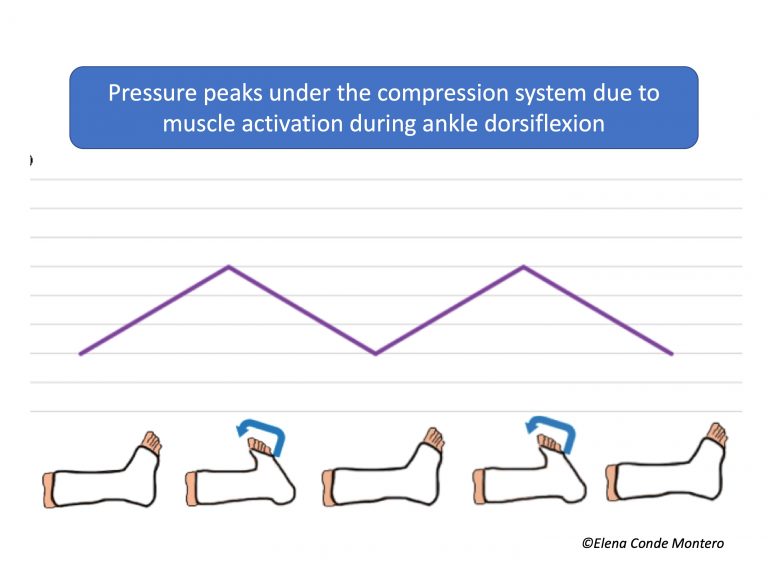
Compression therapy is the mainstay of treatment for venous hypertension, but it should always be associated with physical exercise (adapted to the situation of each person) and anti-edema measures (essentially rest with elevated lower limbs), as we saw in the post “Essential Decalogue in the fight against venous hypertension“. In the blog entry “Phlebolymphedema, a term that should more frequently used” we explained the need for physical exercise to activate the plantar and calf pump, and thus promote venous and lymphatic circulation. As we also commented in the post “Band and bandage: not the same thing“, the higher the stiffness index of the compression system used, the more effective it will be in resisting muscle contraction and achieving high pressure peaks that create brief and intermittent venous occlusions, similar to the physiological functioning of the valves. As muscle contraction is so relevant, muscle atrophy or limited mobility may diminish the effectiveness of compression bandages.

So, if exercise is so important, now that we can’t go for a walk, what can we do at home to encourage venous return?

Young people, with few joint problems and good mobility, can do planned exercises, with or without apparatus (exercise bike), to activate the muscle pump of the calf and foot (let’s not forget that the plantar muscles are very important).

However, being old and having reduced mobility, in addition to other associated comorbidities, does not mean that it is impossible to activate the muscle pump. Here you can find some exercises for dorsal and plantar flexion of the ankle that you can do in a series, for example, of 20 repetitions several times a day, as long as you remember it. The ideal is to do them completely lying on your back, with your legs raised, supported by 2-3 cushions/pillows. If lying in bed is only possible at night, we will take advantage of this moment to do it this way and, the rest of the day, we can do it sitting up, with our legs elevated.

As we said at the beginning, anti-edema measures are fundamental, so we recommend keeping your legs elevated whenever you are sitting. If you have trouble difficulty keeping your legs elevated for a long time, which would be optimal, we can also do the following exercises to activate the muscle pump.

If, in addition to phlebolymphedema and immobility (wheelchair), there is also a certain degree of arteriopathy (ABI>0.5), high  stiffness index bandages with pressures lower than 30 mmHg (or Velcro wraps) is a safe and effective option if the dorsiflexion and plantar flexion exercises we have just seen are performed.

Before finishing, remember that these exercises also help maintain or improve the flexibility of the ankle joint, which is fundamental for the proper functioning of the plantar and calf muscle pump during walking (“Improving the calf pump using home-based exercises for patients with chronic venous disease”).

I am taking this opportunity to send you all a big hug. The wound clinic will be closed for a while, but anything that can be resolved telematically, do not hesitate to consult me. The next few weeks my work will be very different from usual and I will have a new team, made up of exceptional professionals, who are extremely supportive and who give their best every day to help people with COVID-19. Take care of yourselves 🙂

Punch grafting as a painkiller for painful ulcers

Any excuse is always perfect for me to talk about punch grafting. But this time I have a “scientifically important” reason to do it. We have just published an article about the benefit of this technique in painful wounds, and it is the longest series with the greatest variety of aetiologies published so far. Antes de continuar, me gustaría mencionar la gran labor en la recogida y análisis de datos para este estudio que realizó Yasmin de Farias Khayat, en su momento estudiante de último curso de Medicina en Brasil. Nos acordamos mucho de ti…:)

In other enties of the blog we have already talked about the interest of grafting hard-to-heal wounds, despite not having wound beds with ideal conditions, to reduce pain and promote healing, thanks to the growth factors released.

The analgesic effect of punch grafting had already been registered in two previous case series. Fourgeaud et al included 41 patients, with a mean age of 73 years, who mainly presented with venous, arterial and Martorell’s ulcers. Pain was assessed by a numerical scale before, on the day after graft coverage and at discharge. In most patients, basal and breakthrough pain showed significant improvement from the first day after the procedure (75%) and this pain control increased to 90% on the last day in hospital. They also found a reduction in opioid use after the procedure. Nordström et al presented a series of 22 patients with leg and diabetic foot ulcers, all presenting with adequate granulation tissue. The mean score recorded with the visual analogue scale (VAS) for pain was 4.2 before grafting, compared to 0.8 one week later.

Now let’s get on with our series! Although you have the full article at the end of the post, I will make a brief summary:

We conducted a prospective study including 136 patients, with an average age of 60. This is the distribution by ulcer etiolog:

  • 51 venous ulcers (38%)
  • 29 post-surgical ulcers (21%)
  • 15 Martorell ulcers (11%)
  • 15 post-traumatic ulcers (11%)
  • 4 arterial ulcers (3%)
  • 22 ulcers with other causes (16%)

We included patients with and without pain. We measured the pain intensity, with a visual analogue scale (VAS), before punch grafting (t0) and during the the following 3 weeks (t1,t2,t3).  The main outcome was pain reduction.

Here are the main results and conclusions of the study:

  1. Pain reduction was clinically and statistically significant throughout the registered visits in the study.
  2. Patients who presented with non-painful ulcers remained pain-free.
  3. Although pain intensity was only registered before the procedure and during the following 3 weekly visits, many patients reported that pain reduction had started shortly after the procedure, even on the same day.
  4. Unlike the other published series, many of the wounds included in our study did not have a wound bed with optimal conditions for grafting. However, this does not prevent the grafts from having analgesic power, as we found no correlation between the percentage of graft taking and pain reduction.
  5. Punch grafting may help control pain in any type of wound. Although its most widespread use is in venous and Martorell ulcers, as we see in the study, other wounds such as arterial, post-traumatic, post-surgical and atypical ulcers (post-vasculitis or inactive pyoderma gangrenosum) may also benefit from this simple technique.
  6. Although venous ulcers are not classically considered painful wounds, in our series half of patients with venous ulcers had an EVA score for pain >5 . At the time of the first follow-up visit, two-thirds of patients with venous ulcers had achieved complete pain suppression.

Acabo con las dos reflexiones con las que finaliza el artículo:

  • ¿Cuáles son las barreras que impiden que la técnica del injerto en sello  se extienda entre los profesionales sanitarios.
  • ¿Cuál es el mecanismo exacto por el que los injertos en sello disminuyen el dolor independientemente del porcentaje de prendimiento?
injertos en sello heridas dolorosas

Bandages impregnated with zinc oxide: Unna boot and much more

Unna boot is not something new. In fact, it’s something very old (almost as old as punch grafting:). I was waiting for the slightest excuse to write about this type of bandage and take the opportunity to comment on the versatility of the use of zinc oxide impregnated bandages… The truth is that scientific publications on this subject are not frequent, so, as an article on the history of this bandage1 and a literature review protocol2 have just been published, I already have a “current” excuse. If we add to this that our team has talked about zinc impregnated bandages as a therapeutic pearl in several communications and posters of the congress of the Sociedad Española de Heridas (SEHER) that, one more year, has just taken place in Madrid; it is mandatory to talk about zinc oxide again in this blog!

What is exactly the Unna boot bandage?

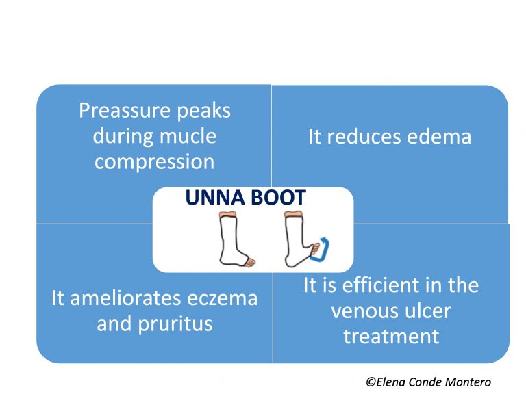
It is a compression system made up of inelastic gauze bandages impregnated with zinc oxide. Therefore, it is a bandage with a high stiffness index, which exerts pressure peaks during the calf muscular contraction, with the consequent reduction of ambulatory venous hypertension (See post “Band and bandage: not the same thing”). In other words, it is an interesting alternative for the treatment of venous ulcers in active patients. The pressure exerted will vary depending on the number and overlapping layers of bandage, as well as the type of secondary bandage applied over it.

But the benefit of this bandage is not limited to its stiffness, but the fact that it is impregnated with zinc oxide makes it especially interesting. As we commented in the post “Why do we use topical zinc on wounds and perilesional skin?”, zinc oxide has anti-inflammatory and epithelialization promoting properties, very useful in cases of stasis eczema. Different models of this type of bandage have been commercialized, with variability in the concentration of zinc oxide and other moisturizing components, such as glycerin. It is a wet bandage, which dries and hardens on the patient’s skin, without limiting ankle joint movement.

Why is it called Unna boot?

Because it was invented by the German doctor Paul Gerson Unna (1850-1929). This doctor is a very relevant figure in the history of medicine, especially for his interest and research in skin diseases, such as dermatitis. As an alternative to the poorly tolerated compression bandages of his time, in 1885 he invented a gauze bandage impregnated with a mixture of 15% zinc oxide in a glycerin and gelatin-based paste. This mixture had drying and “cooling” effects, as well as anti-pruritic action due to its continuous and slight pressure on the skin. After numerous tests in the following years, Unna presented a new form of compression bandage at the Third International Congress of Dermatology in London in August 1896 and published it that same year. His bandaging technique later became widely recognized for its effectiveness and simplicity.1

What do the studies say about its usefulness?

The studies say FEW THINGS, because they are few, with few patients and very few have a control group… As I commented at the beginning of the post, a group of Brazilian professionals has designed and published a protocol to make an exhaustive review on the subject.2 It is not surprising that they are experts from Brazil, since most of the articles that we find in the literature are signed by Brazilian authors (it is undoubtedly a widespread technique in this country).

The most recent review on Unna boot, published in 2018, highlights the scarcity and low quality of the studies published (14 papers), mostly cases and case series.3

In this context and despite the fact that the available evidence supports and recommends the use of multi-component compression systems, the Unna boot is an economical and efficient alternative in certain countries and for a certain patient profile, especially those active people who present significant eczema associated with venous ulcers.

Practical questions about the use of the Unna boot

I’m sure that you’re having a lot of questions about how to use this bandage, so here you will find some practical recommendations:

  1. The frequency of dressing changes will vary depending on the wound exudate, reduction of edema and tolerance to the progressive dryness of the bandage on the skin, but we could keep it on for a week.
  2. We will use a secondary bandage, whose purpose may be only to protect/fix or also to add pressure. Depending on the objective we are looking for, we will select the type of bandage. This secondary bandage will help to avoid the rapid fall of the Unna boot after the reduction of the edema.
  3. In cases of eczema, the application of topical corticosteroids prior to the placement of the bandage will reinforce and increase the anti-inflammatory effect.
  4. The zinc bandage can be the primary dressing for the wound, whose exudate we will control with secondary dressings between the zinc bandage and the secondary bandage.
  5. It is necessary to avoid folds, because when dry, they will be potential areas of friction and the occurrence of new wounds.
  6. In older patients with little mobility or thin legs and risk of hyperpressure on bony prominences, associated arteriopathy or lack of sensitivity due to neuropathy, we will not indicate Unna’s boot. In these cases, we will use another type of bandage, for example multi-component bandages, always with protective padding, adapted to the person’s needs.

Beyond Unna boot

Perhaps the “point 6” has discouraged many of you, since this type of patient I describe is very common in the practice… But, as the title of this entry says, zinc-impregnated bandages are not limited to Unna boot! In fact, we often use them as a dressing, primary or secondary, mainly when we have associated eczema (usually in combination with topical corticosteroids). The drying effect of zinc oxide is very useful in case of acute exudative eczema.

To increase the concentration of zinc, we can add zinc oxide in solution or cream. It is a great alternative to protect and treat perilesional skin and very interesting in the wound bed (especially after covering with punch grafting:). During dressing changes,we will avoid cleansing by rubbing to eliminate the residual zinc oxide in the skin since it is not necessary to remove it and, in addition, it would worsen the eczema.

In patients who do not have an active ulcer but do have eczema secondary to venous hypertension, we sometimes recommend that they apply a  coricosteroid cream and cover it with a piece of zinc bandage under their compression stocking or self-adjusting Velcro device.

Used as a dressing or bandage, our experience confirms that the zinc impregnated bandage has an important anti-inflammatory action in different leg dermatitis, normally combined with topical corticosteroids, achieving great symptomatic relief. Besides leg eczema, its use in pustulosis and erosive dermatosis is also interesting4 (See post: “: “Pustular and erosive leg dermatosis: you may have seen a case without knowing it “).

“When knowledge that is generated in team is shared in team, the passion with which it is transmitted is exponentially multiplied”. This is what  pur team feel whenever we are all at a congress 🙂 Of course, I am also very excited when talking about wounds with my dermatologist friends!


Referencias:

  1. Tekiner H, Karamanou M. The Unna Boot: A Historical Dressing for Varicose Ulcers. Acta Dermatovenerol Croat. 2019 Dec;27(4):273-274.
  2. Saidel MGB, Oliveira HC, Dini AP, Kumakura ARSO, Melo Lima MH. Assessment of the use of Unna boot in the treatment of chronic venous leg ulcers in adults:systematic review protocol. BMJ Open. 2019 Dec 23;9(12):e03209
  3. Cardoso LV, Godoy JMP, Godoy MFG, Czorny RCN. Compression therapy: Unna boot applied to venous injuries: an integrative review of the literature. Rev Esc Enferm USP. 2018 Nov 29;52:e03394.
  4. Di Altobrando A, Patrizi A, Vara G, Merli Y, Bianchi T. Topical zinc oxide: an effective treatment option for erosive pustular dermatosis of the leg. Br J Dermatol. 2020 Feb;182(2):495-497.

Iron, ochre dermatitis and venous disease

The idea of talking about iron and venous ulcer came to me when I read an article that has just been published on the potential relationship between iron deficiency and venous leg ulcer, suggesting the possible role of iron loss from wound exudate:Does localized iron loss in venous disease lead to systemic iron deficiency? A descriptive pilot study1. In addition, the authors of this paper have also just published a review on iron deposits, anaemia and venous ulcers:An overview of the relationship between anaemia, iron, and venous leg ulcers “2

Since there is still much to be researched on the iron-deficit/venous ulcer relationship and the actual impact of anemia on healing, let’s talk about a topic that has been studied in more detail, although the available evidence is not very robust either: The role of local iron deposits in the evolution of chronic venous insufficiency.

Which functions has iron in the body?

Iron is the most abundant trace element in our organism and plays a fundamental role in many cellular functions. It is an essential part of haemoglobin as it facilitates the transport of oxygen to tissues. It participates in multiple metabolic processes, since it is part of enzymes and other molecular complexes. In addition, it also has an important function in tissues with a lot of cell turnover, such as skin, since it regulates DNA replication.

Despite being a fundamental metal for life, in high quantities it can be toxic. This is because free iron has the ability to yield or donate electrons easily (cyclic shift from the Fe2+ to Fe3+ state) and can catalyze free radical reactions and increase oxidative stress.

Our body is responsible for limiting this potential damage by storing iron in protein complexes, such as haemoglobin, transferrin or ferritin. In addition, there are many regulatory mechanisms to maintain an adequate balance between free (hazardous) and protein bound iron. However, these regulatory mechanisms are not sufficient when there is uncontrolled iron overload, both at systemic and local levels.

Why do cutaneous iron deposits occur on the legs?

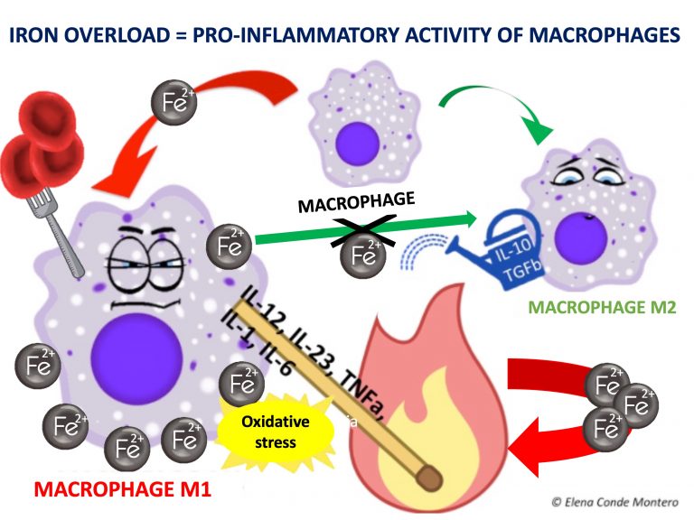
Uncontrolled venous hypertension, which causes chronic venous insufficiency (CVI), leads to extravasation of red blood cells into the skin. These extravasated red blood cells are ingested and degraded by macrophages in the dermis, which release and store iron inside them, first in the form of ferritin and then, when saturated, in the protein complex called hemosiderin. These iron deposits, in addition to melanin deposits (melanocytes are activated by inflammatory processes affecting the skin, such as CVI), produce a brownish coloration in the skin, which is called ochre dermatitis and is one of the clinical characteristics of stage C4 of the CEAP classification of chronic venous disease (See post “CEAP classification of chronic venous disorders: let’s all speak the same language“). What is the weight, separately, of haemosiderin and melanin in the ochre colour of these legs? This is a difficult question to answer, since hemosiderin activates melanin production by melanocytes…

The problem of this hyperpigmentation is not only aesthetic. Iron deposits in the skin seem to have great importance in the evolution of the venous disease and, therefore, of the venous ulcer. In fact, in our clinical practice we observe that leg ulcers with extensive deposits of hemosiderin are more resistant to treatment.

What has been researched on hemosiderin deposits and the evolution of venous disease?

Published studies are scarce and include few patients. Caggiati’s group performed 2 studies in which they analysed haemosiderin deposits in skin biopsies of legs in different stages of chronic venous disease.3,4 In both studies, most patients with normal skin colour did not have haemosiderin deposits in the biopsy (detected by Perls staining). In patients with hyperpigmentation (C4), haemosiderin deposits were only found in those with higher pigmentation. In fact, in the initial stages, these authors associate brownish colour with a greater amount of melanin, since, as we have said, melanocytes can be activated in all cutaneous inflammatory processes. In more advanced stages, such as lipodermatosclerosis (C4b) and venous ulcer (C6) these deposits were always found. Patients with healed ulcers (C5) were not included, so no conclusions can be drawn from this group. The authors conclude that, despite not being able to draw physiopathological conclusions, it is clear that the progressive skin changes of chronic venous disease are associated with the presence of haemosiderin deposits. They also suggest that there may be some genetic condition that justifies a greater deposit of iron (and also melanin) in certain patients.

Regarding to the few studies comparing iron levels in chronic vs. acute wound exudate and in the edges vs. perilesional tissue, the variability between the methods used for detection stands out. However, the results point to a higher amount of iron in wound bed and edge of chronic ulcers.

What role may iron deposits play in the chronicity of wounds?

The differentiation of macrophages and their passage from M1 (pro-inflammatory) to M2 (anti-inflammatory) type is fundamental in the transition from the inflammatory to the proliferative phase of the healing process. Iron seems to be involved in these changes since, when ingested by M1, it maintains its activity and hinders its transformation into M2.

In other words, iron deposits favour the persistence of M1 macrophages and the consequent release of pro-inflammatory cytokines. In addition, free oxygen radicals released by the chemical reactions of iron ions in macrophages cause the accumulation of senescent fibroblasts resistant to apoptosis, which also release pro-inflammatory factors.5 This pro-inflammatory microenvironment produces more blood extravasation and, therefore, more iron deposits, producing a vicious circle.

What could be done to control the pro-inflammatory action of iron deposits?

The most reasonable would be to look for a method of chelating (sequestering) iron within the wound to prevent the release of reactive oxygen species by the ionic iron. Deferoxamine is an established iron chelator as a systemic treatment for iron overload situations such as hemochromatosis.

Would a deferoxamine-impregnated dressing be useful? More than 15 years ago, specific in vitro studies were published that pointed to the interest of this chelator applied topically…6,7Hmmm Why has no further research been done in this line?

Hopefully further studies will be carried out to find out the real role of these iron deposits in the chronicity of venous ulcers and, therefore, their potential role as a therapeutic target!

Referencias:

  1. Ferris AE, Harding KG. Does localized iron loss in venous disease lead to systemic iron deficiency? A descriptive pilot study. Wound Repair Regen. 2020 Jan;28(1):33-38.
  2. Ferris AE, Harding KG. An overview of the relationship between anaemia, iron, and venous leg ulcers. Int Wound J. 2019 Dec;16(6):1323-1329.
  3. Caggiati A, Rosi C, Casini A, et al. Skin iron deposition characterises lipodermatosclerosis and leg ulcer. Eur J Vasc Endovasc Surg. 2010;40(6):777-782.
  4. Caggiati A, Rosi C, Franceschini M, Innocenzi D. The nature of skin pigmentations in chronic venous insufficiency: a preliminary report. Eur J Vasc Endovasc Surg. 2008;35(1):111-118.
  5. Wlaschek M, Singh K, Sindrilaru A, Crisan D, Scharffetter-Kochanek K. Iron and iron-dependent reactive oxygen species in the regulation of macrophages and fibroblasts in non-healing chronic wounds. Free Radic Biol Med. 2019 Mar;133:262-275.
  6. Wenk J, Foitzik A, Achterberg V, et al. Selective pick-up of increased iron by deferoxamine-coupled cellulose abrogates the iron-driven induction of matrix-degrading metalloproteinase 1 and lipid peroxidation in human dermal fibroblasts in vitro: a new dressing concept. J Invest Dermatol. 2001;116(6):833-839.
  7. Taylor JE, Laity PR, Hicks J, et al. Extent of iron pick-up in deforoxamine-coupled polyurethane materials for therapy of chronic wounds. Biomaterials. 2005;26(30):6024-6033.


Leg wrinkles are beautiful” and other thoughts in the 5 year blog anniversary

Five years of the blog “When a wound in the skin won´t heal also means five years of “We have to think and do anything to close it as soon as possible. It could not be otherwise because the story of this website is closely related to that of the wound clinic in Centro de Especialidades Vicente Soldevilla, Madrid. It all started in 2014 with Soledad Guisado, my dearest Sole, who trusted and supported me from the very first moment.

Nobody said the beginnings were easy, but we survived and since then the team has grown more and more. It has changed, too. But change is a purely formal issue because, sentimentally, all of you who have been part of this project at some point, will always remain part of the team 🙂

The great novelty of the last year has been the alliance of collaboration that we have established with the Department of Angiology and Vascular Surgery of the Hospital de la Cruz Roja, thanks to the motivation of its head of Service, the great expert in Phlebology (and beautiful person) Lourdes Reina.

If there is one thing that I have no doubt about (and time confirms it) it is that “in our wound clinic we can help a few people, but, by sharing knowledge and experiences, that help may increase exponentially”. This is the main idea of the blog, for which it was born and for which it is being maintained. But it is also my philosophy when organizing and participating in training activities.

In this context, two weeks ago we organized in our clinic a training  course on leg ulcers for professionals who work both in primary care and in nursing homes, with the collaboration of  La Roche Posay. We talked about the importance of a correct diagnosis for a successful treatment, the theoretical and practical principles of compression therapy and, as it could not be otherwise in a course conducted by our team, our star technique, punch grafting. The training of these professionals and the establishment of pathways to collaborate with them is essential. Essential because MOST PEOPLE WITH LEG LEGS ULCERS ARE CARED FOR IN PRIMARY CARE AND RESIDENCES FOR THE ELDERLY, NOT IN SPECIALIZED CARE CENTERS. Let’s get out of our offices and go where the problem is!


And many times the problem has started as a simple “wound” after a silly blow to the leg, which has grown over months into a large and extremely painful ulcer that has disrupted the life of an elderly person… On numerous occasions, the trigger is a minor laceration Although we usually make the mental association “uncomplicated traumatic wound” = “acute wound” = “this closes smoothly without doing anything”, when the location is the leg, even in healthy patients without venous disease, these wounds can take time to close due to the pro-inflammatory effect of the force of gravity, which generates a state of venous hypertension. If the person has chronic venous insufficiency, the inflammation in the wound will be even greater. The issue is more complex  in older people, whose skin fragility and age-related vascular damage (even without other comorbidities) are already a big reason to think about “potential chronic wound if we do not perform an adequate anti-inflammatory strategy“. And what does “anti-inflammatory strategy” mean in these patients? Essentially therapeutic compression and anti-oedema measures, which is the best anti-gravity and anti-inflammatory strategy for any leg ulcer provided there are no contraindications. See post “Compression is the key to treating leg wounds”. In addition, we will also select the dressing that best suits the conditions of the wound bed, protecting and preserving the perilesional skin.

Therapeutic compression adapted to the patient’s needs (always after pulses palpation and, in case of doubt, ABI greater than 0.5) and the recommendation of anti-oedema measures (walking, resting with the legs elevated, foot dorsiflexion exercises) can and should be done immediately after the occurrence of the wound, both in primary care and in nursing homes. Regarding compression bandages, we must insist on the protective padding, to avoid areas of hyperpressure in these fragile skins (see post “Wounds secondary to dermatoporosis or chronic cutaneous insufficiency” and “Band and bandage: not the same thing“). The degree of pressure must be adapted to the possible arteriopathy and other comorbidities of the patients. See post “Compression is the key to treating leg wounds”.

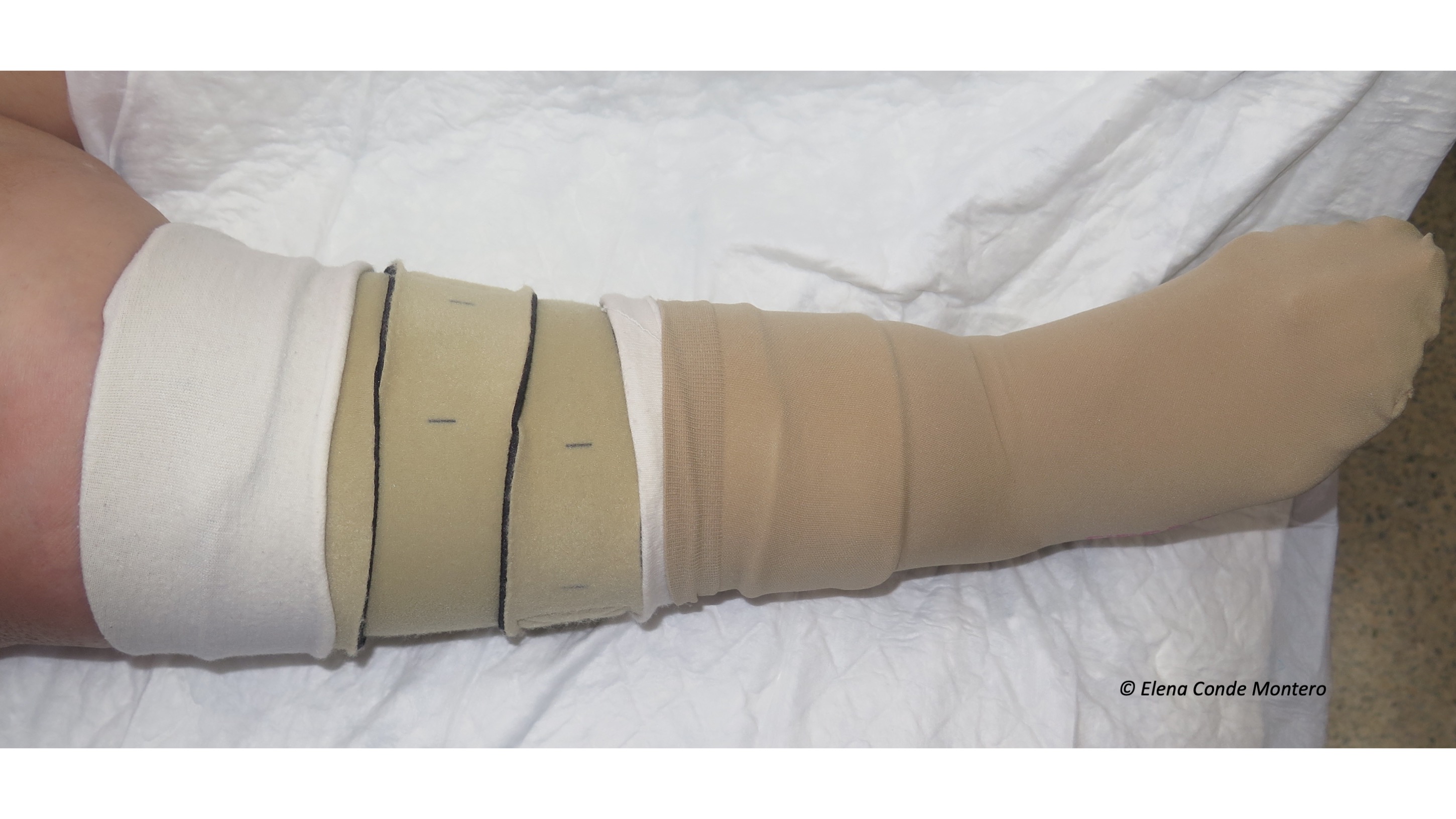
Compression stockings and adjustable compression wrap devices are interesting alternatives, mainly in the case of small wounds, patient preferences or inability to change bandages as often as necessary. In legs with dermatoporosis it is essential to use protective socks under the stocking (or wrap device) to avoid friction damage to the perilesional skin.

This is an example of a patient with skin tears treated in the first 48 hours with the best anti-inflammatory strategy. We adore to see these big wrinkles when we remove the compressive bandage, a sign of its effectiveness in reducing oedema…And these leg wrinkles are the most beautiful ones 🙂

Speaking of beauty, we can not fail to mention the beautiful mosaics that form punch grafts in the wound bed. As we saw in the postLarge leg wounds after mild trauma“, early punch grafting is the solution in many of these post-traumatic wounds to slow their progression, reduce pain and promote epithelialization. A simple solution, very well accepted and very efficient for a terribly limiting problem! 

The success is in acting as soon as possible. Not only when the wounds have occurred, but already with preventive initiatives. Moisturized legs, with good control of edema and covered with protective tissues and devices to avoid potential trauma, will have fewer problems. Fluid communication between professionals working in nursing homes, primary care and specialized care is the key in preventing chronic wounds from developin in the elderly. TOGETHER WE CAN CHANGE THE LIFE OF THESE PEOPLE, REDUCING THEIR DAYS SUFFERING WITH WOUNDS!

Happy New Year and thank you very much for your confidence and interest in the blog! I hope to keep writing and drawing with the same excitement and love for many more years 🙂

Vitamins, folic acid, proteins and venous ulcer

  • What would you include in a blood test for a patient with a venous ulcer?
  • What alterations would you expect to find, whose adjustment may have a direct impact on wound healing?

This post aims to provide guidance on these questions, the answers to which, at present, are not agreed upon. Let’s see what the published studies say about this!

The idea of writing a post on this topic came to me when I read the article published last month “Nutrition status in patients with wounds: a cross-sectional analysis of 50 patients with chronic leg ulcers or acute wounds”. Among the authors of the study, in addition to dermatologists and internists, we find a psychologist, an essential specialist, as we will see, to address the nutritional problems of people with leg ulcers.

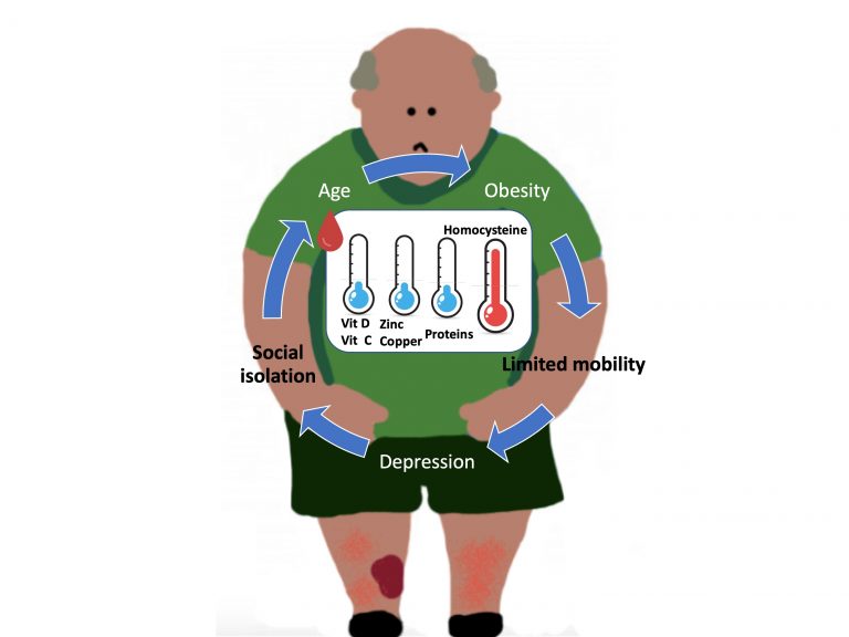
When we talk about nutrition and wounds, the first thing we always think about, and in fact this is what has been mostly studied, is the relationship between nutritional deficit and poor evolution of pressure ulcers. However, this idea of a nutritional approach should be integrated into the therapeutic strategy of any wound, especially in elderly patients. In fact, age, with the associated decrease in taste and appetite, as well as problems with chewing, is a major risk factor for malnutrition. Malnutrition results not only from low calorie intake, but also from a lack of micronutrients. In other words, obesity, which is so common in our patients with leg wounds, can be accompanied by a nutritional deficit due to a diet poor in vitamins and essential amino acids. Patients who live alone, suffer from depression or have little mobility for shopping and preparing food are also at risk of malnutrition.

Interestingly, there is an overlap of risk factors for malnutrition and venous ulcers: age, obesity, social isolation, depression, poor mobility. And it is in a vicious circle, so a psychosocial approach is essential for the holistic treatment of these patients.1

Well, coming back to our patient with a venous leg ulcer, what alterations should I look for in his blood test to treat them and improve wound healing?

I can tell you that, at this moment, there is no consensus on what nutritional parameters to ask for, nor if, in the event of finding alterations in their levels, dietary supplements have an impact on wound healing But let’s see what has been studied about this… First of all, I would like to stress that no association has been found between malnutrition and the size or duration of the leg ulcer.1,2

VITAMINS

  • VITAMIN C

Its deficit is frequent in patients with leg ulcers. This vitamin is fundamental in the inflammatory phase, since it is involved in the apoptosis of neutrophils, and in the proliferative phase, since it participates in the synthesis of collagen. Clinical trials are needed to detect the usefulness of its supplementation in leg wound healing.1,2

  • VITAMIN D

Its deficit is frequent in the general population. In fact, in studies with a control group, such as the one we indicated at the beginning of the post, this deficit is found in both groups, even though it is more frequent in the group with leg ulcers. This may be due to the reduced mobility of these patients, with less exposure to sunlight. The anti-inflammatory effect of the vitamin may be of interest in modifying the pro-inflammatory environment of chronic ulcers. A clinical trial compared supplementation with 50,000 IU per week of vitamin D for 2 months with placebo in patients with venous ulcers and a deficit of vitamin D. Despite finding a trend towards greater healing rates in the treatment group, the differences are not significant, the number of patients included is small and the variability of lesion sizes is very high .

TRACE ELEMENTS

  • ZINC

This trace element was the main character of a previous post (“Why do we use topical zinc on wounds and perilesional skin?”)… In fact, in its topical presentation it is one of the main treatments used in our clinic... However, while its topical use is widespread, the available evidence does not support the interest in healing of oral zinc administration, even if there is a deficit of it.1 It must be taken into account that clinical trials are scarce, of small size and multiple limitations, as highlighted in the conclusions of the last Cochrane review on this subject.4

PROTEINS

While protein supplements are a well-established strategy in pressure ulcers, in venous ulcers this recommendation does not exist, even though many of these patients have hypoproteinaemia. The most common measure for testing blood protein levels is serum albumin concentration. This protein deficit is thought to be associated with the catabolic state of having an ulcer, due to increased metabolic demand, rather than protein loss in the exudate.1,5

However, there is one amino acid that is frequently elevated in patients with leg ulcers:

  • HOMOCYSTEINE

Homocysteine is a sulfur-derived amino acid derived from methionine, in whose metabolism several enzymes are involved. It has a cytotoxic action on the endothelium, increasing pro-inflammatory factors and oxidative stress. It also facilitates platelet adhesion, hence its potential thrombogenic effect. Its high levels in blood, hyperhomocysteinemia, is considered an independent risk factor for arterial and venous thrombosis, and is usually related to old age, renal failure, genetic and nutritional factors, in addition to being a marker of tissue damage. Considering this microthrombosis-enhancing action, it is easy to think that homocysteine is involved in the development of venous ulcer, but it should not be forgotten that it can also simply be a marker of tissue damage…6

Studies comparing blood homocysteine levels in patients with chronic venous insufficiency, with and without ulcers, with a control group always detect a higher concentration in patients in the first group, with statistically significant difference. Furthermore, there seems to be no correlation between the evolutionary stage of chronic venous insufficiency and hyperhomocysteinaemia. A study that included arterial, venous and mixed leg injuries also found no association of hyperhomocysteinemia with any of these particular etiologies.6

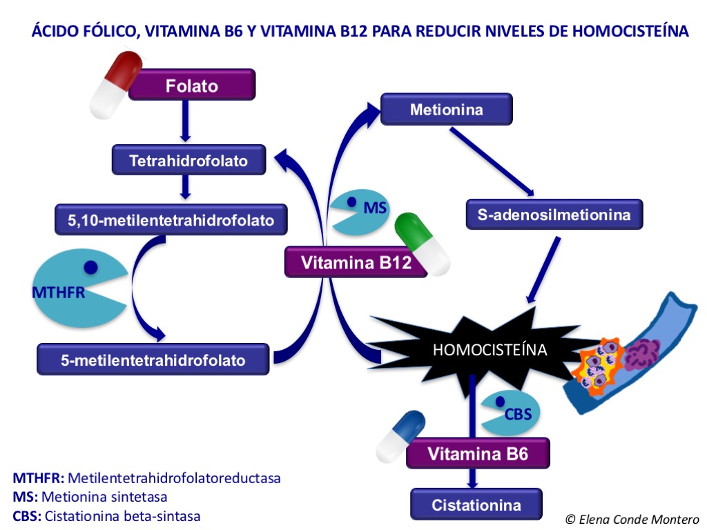
As we discussed in the post “Ulcer secondary to alteration of homocysteine metabolism mimicking pyoderma gangrenosum”, the treatment of hyperhomocysteinemia is very simple, since the contribution of folic acid facilitates the conversion of homocysteine into methionine. This scheme on homocysteine methionine metabolism also helps us understand why vitamin B6 and B12 would be interesting as well.

So, is there a causal relationship between leg ulcer and hyperhomocysteinemia? Is hyperhomocysteinemia a cause or a consequence? Does lowering the homocysteine concentration have an impact on leg wound healing?

Although different studies have been conducted to analyse the prevalence of hyperhomocysteinemia in patients with leg ulcers, none has been designed with the aim of studying its potential causal relationship. Therefore, we know that hyperhomocysteinemia is common in leg ulcer patients but we do not know exactly why.6

Regarding the benefit of treating hyperhomocysteinemia on wound healing, more studies are needed. What does seem clear is that reducing hyperhomocysteinemia does not reduce cardiovascular and thromboembolic risk.6 One clinical trial examining the value of daily folic acid supplementation for 1 year in patients with leg ulcers and hyperhomocysteinemia found statistically significant differences in the healing rate in favor of the treated group. It is a safe and simple treatment, so although there is not much evidence and at the moment we do not know if the elevated homocysteine is a cause or a consequence, I think it is interesting to start the treatment. 

As we have just seen, the available evidence doesn’t make it clear at all which deficits have a direct impact on healing and which nutritional supplements are useful in people with leg ulcers. In fact, it would be also interesting to conduct studies in order to determine whether these nutritional supplements are interesting in patients with normal serum values… The benefit of these supplements might be only achieved at higher doses than those considered normal… What an unresolved dilemma! What is clear is that we have to recommend (besides explaining) is a healthy diet.

My friend Nicolas Kluger, dermatologist and artist, explains it to us with a watercolor 🙂 Je te remercie pour ta belle peinture, Nicolas!

I would like to finish with a “NUTRIPSICOSOCIAL” reflection: Considering that obesity, lack of mobility and depression are modifiable risk factors for malnutrition (age unfortunately is not), let us try to modify them to improve the nutritional status of people with leg ulcers!

References:

  1. Renner R, Garibaldi MDS, Benson S, Ronicke M, Erfurt-Berge C. Nutrition status in patients with wounds: a cross-sectional analysis of 50 patients with chronic leg ulcers or acute wounds. Eur J Dermatol. 2019 Dec 1;29(6):619-626.
  2. Barber GA, Weller CD, Gibson SJ. Effects and associations of nutrition in patients with venous leg ulcers: A systematic review. J Adv Nurs. 2018;74:774– 787.
  3. Burkiewicz, C. J. C. C., Guadagnin, F. A., Skare, T. L., Do Nascimento, M. M., Servin, S. C. N., & de Souza, G. D. (2012). Vitamin D and skin repair: A prospective, double-blind and placebo controlled study in the healing of leg ulcers. Revista do Colegio.
  4. Wilkinson Ewan, A. J. Oral zinc for arterial and venous leg ulcers. Cochrane Database of Systematic Reviews. 2012.
  5. Legendre, C., Debure, C., Meaume, S., Lok, C., Golmard, J. L., & Senet, P. (2008). Impact of protein deficiency on venous ulcer healing. Journal Of Vascular Surgery, 48, 688–693.
  6. Studer M, Barbaud A, Truchetet F, N’guyen PL, Bursztejn AC, Schmutz JL.[Hyperhomocysteinemia and leg ulcers: A prospective study of 68 patients]. Ann Dermatol Venereol. 2011 Oct; 138(10):645-51.
  7. de Franciscis, S., de Sarro, G., Longo, P., Buffone, G., Molinari, V., Stillitano, D. M.,Serra, R. Hyperhomocysteinaemia and chronic venous ulcers. International Wound Journal. 2015;12: 22–26.


Why are adjustable compression wrap devices so popular?

This is another post to add to the list of blog entries on Compression Therapy! And it couldn’t be any other way because it’s the must-see theme this month, when we’re celebrating #YestoCompression. Once again, I was delighted to participate in the European Venous Ulcer and Compression Days. I remember very fondly the enthusiasm with which I prepared the first event of #Yestocompression, in December 2016, and I love to see that this desire for change only grows, and it grows faster in TEAMJ

#YesToCompression is enormously relevant because when faced with a venous ulcer WITHOUT COMPRESSION THERE IS NO CHOICE! Furthermore, the SUCCESS OF COMPRESSION falls with its COMPRESSION. Both the practitioner and the patient should understand it. DIALOGUE with the patient and their caregivers is fundamental for them to understand the NEED for compression therapy in the treatment of their venous hypertension. VENOUS ULCER = VENOUS HYPERTENSION.  But the benefit of compression is not limited to venous leg ulcers: IF THERE IS NO CONTRAINDICATION, COMPRESSION THERAPY IS THE BEST ANTI-GRAVITY AND ANTI-INFLAMMATION TREATMENT FOR ANY LEG ULCER!

Now back to the title of this post. Surely the question many of you will ask yourselves is not why we like them so much, but what kind of compression are they? All of you who are asking yourselves this second question are about to discover a treatment alternative that will solve many of your problems.

What are adjustable compression wrap devices with Velcro-type fasteners?

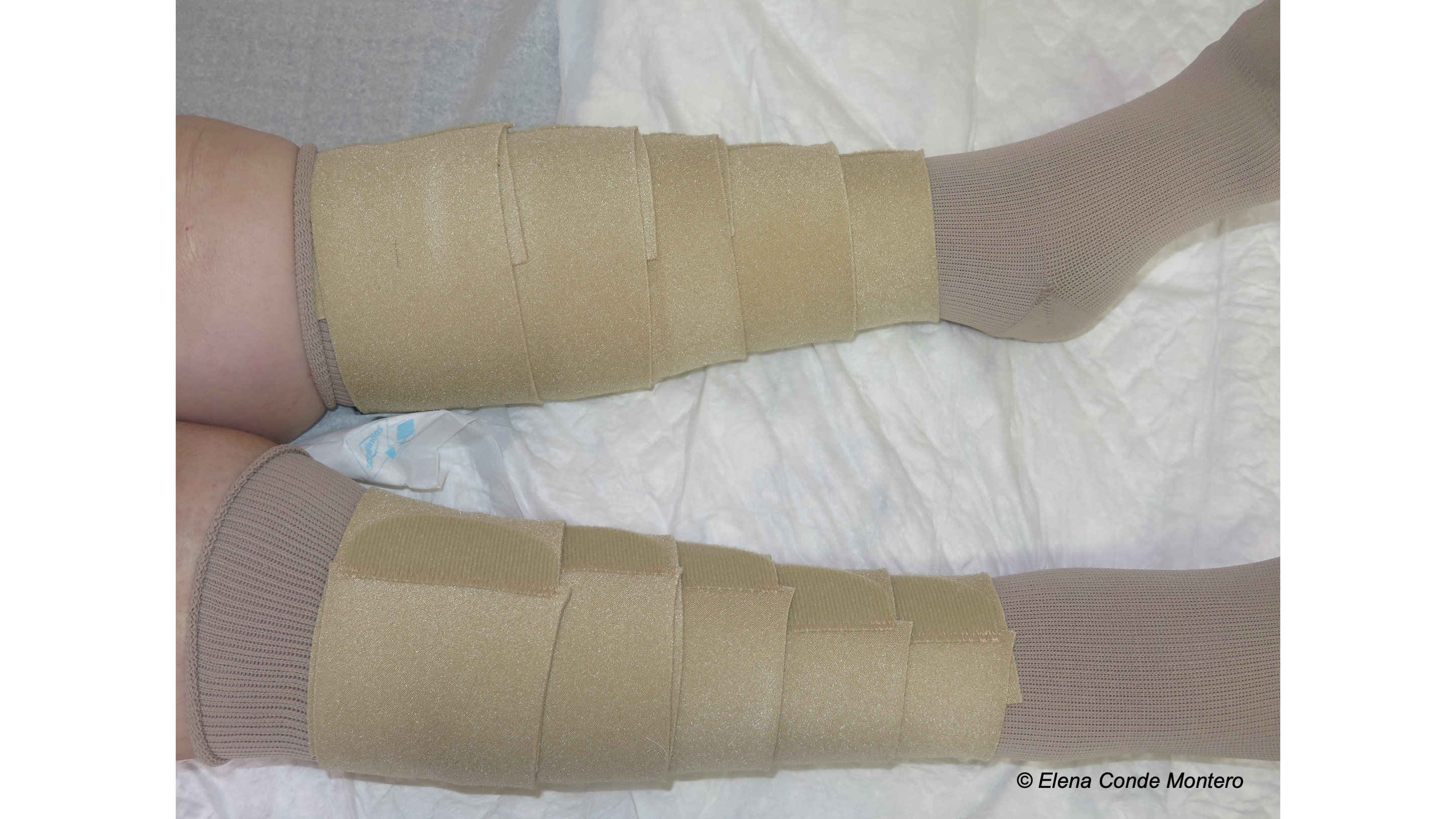
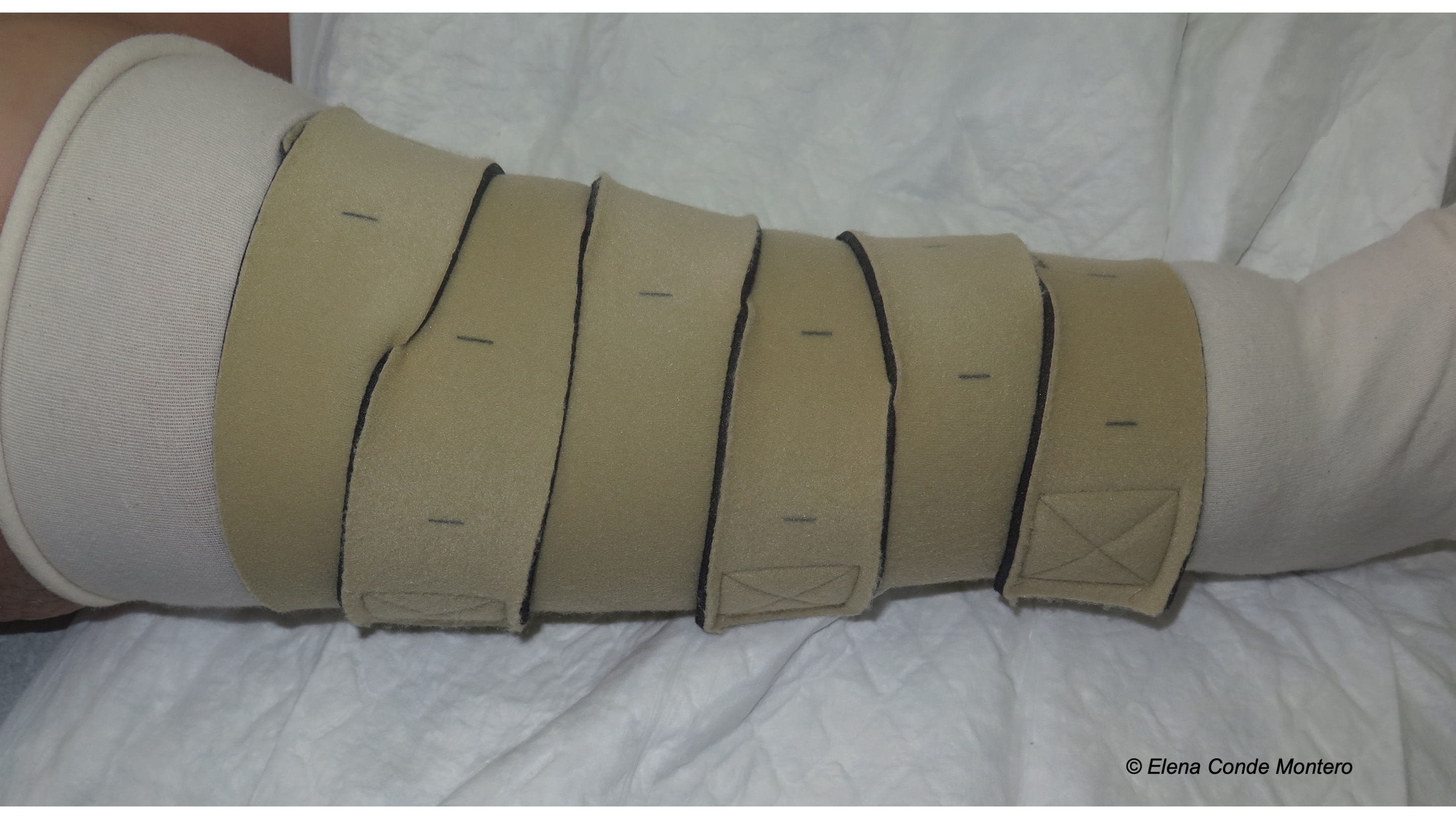
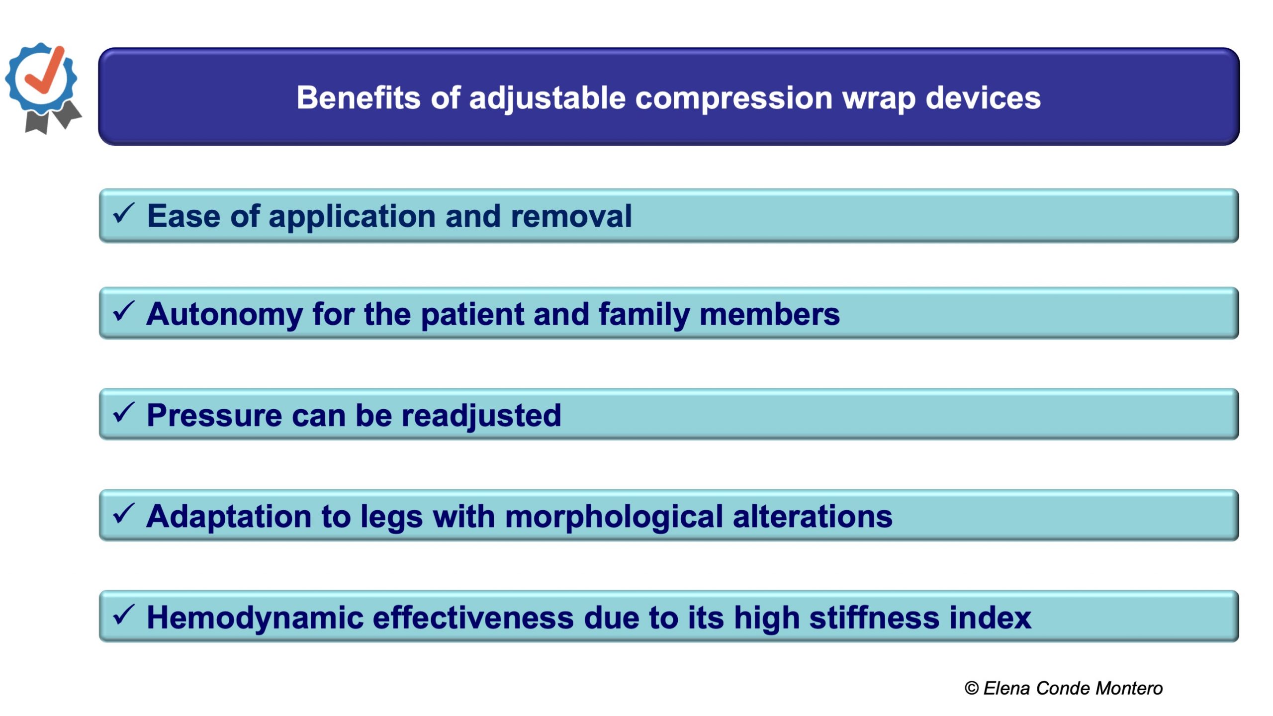
They are one of the most remarkable innovations in the field of compression therapy in recent years. They are garments made of short stretch fabrics, which wrap around the leg and adapt to the shape of the limb, are adjusted according to the desired pressure and are fixed with velcro-type closures. Since the fabric is short stretch, when placed on the leg behaves with a high rigidity index, so it presents resistance to changes in volume in the calf and achieves more effective pressure peaks during muscle contraction than compression stockings, whose fabric is elastic (see post: “Bandage and dressing: not the same thing“).

Moreover, considering that they exert low pressure at rest, they are also usually better tolerated than elastic fabrics.

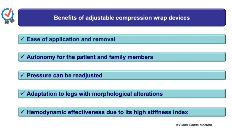
Besides being a system of easy application and removal by the patient himself, it also allows readjustment of the pressure exerted throughout the day, by repositioning the Velcro-type closure. This possibility of adjusting the pressure adapted to the changing volume of the leg avoids the main limitation of short stretch bandages, which is the rapid fall of the bandage associated with edema reduction.

Different designs have been marketed, with variability in the degree of tissue stretching and type of placement. Depending on the model, the system of selecting and controlling the pressure we wish to achieve changes. We always place a protective sock or tubular tissue on the skin before applying the device, which should go from the ankle to below the popliteal fossa. To avoid or control the edema in the foot, we will use a sock or stocking with compression mainly limited to the foot, if it is not enough, we will add a compression system with Velcro limited to the foot. 

When should you use compression wrap devices instead of a bandage or a medical compression stocking?

All three are valid types of compression therapy. The ideal one for each patient will be the one that best suits their characteristics, as well as those of their social environment. Although it can be considered as an alternative to bandaging during the process of wound closure, its main general use occurs when there is no possibility for the professional to perform the dressings as often as necessary or for the patient’s comfort, and it is also an alternative to compression stockings for edema control and prevention of venous ulcer recurrence.

Patients who benefit most from these systems are those who are unable to wear stockings (especially due to obesity or age-related osteoarticular problems), those with extreme or very irregular leg diameters, and those with skin disorders secondary to lymphedema and phlebolymphedema, in which they are a very practical alternative to flat-knit stockings (see post “Phlebolymphedema, a term that should be more frequently used”).

These systems are also an excellent option for patients with chronic venous insufficiency and some degree of peripheral artery disease, who may have poor tolerance of elastic tissues due to their high pressure at rest. In these patients, the application of wrap devices at less than 30 mmHg is not only safe and well tolerated, but, by reducing edema, it also improves arterial perfusion (see post: “Compression is key to treating leg wounds“).

What do the studies say about wrap devices?

Different studies show the ease of application, without the need for special prior training, and the possibility of obtaining high pressures during exercise (also with dorsiflexion movement of the foot), similar to those of a rigid bandage with inelastic bandages, with the added benefit of being able to readjust the pressure.1

A review has just been published on the interest of these Velcro wrap systems in the treatment of venous ulcers. This descriptive review includes 16 studies,2 mostly case series, with very small samples (a total of 192 patients included when adding up all the studies in the review). The only randomised clinical trial (small, 12 patients, and with many limitations), which includes bilateral ulcers and compares the effect of the multi-component bandage on one leg with the wrap device on the other, found a higher healing rate when using the second device.3

The results of the studies in the review suggest the potential improvement in time to healing, reduction in costs and in the number and duration of nursing consultations, and improvement in patients’ quality of life.

Although the available evidence is still very limited, the results of the published studies point to a clear benefit of these devices in achieving adequate therapeutic compression adapted to the patient´s needs.

LET’S ALWAYS BET FOR THE MAXIMUM AUTONOMY AND QUALITY OF LIFE OF THE PATIENTS!

References:

  1. Partsch H. Reliable self-application of short stretch leg compression: Pressure measurements under self-applied, adjustable compression wraps.Phlebology. 2019 Apr;34(3):208-213.
  2. Stather PW, Petty C, Howard AQ. Review of adjustable velcro wrap devices for venous ulceration. Int Wound J. 2019 Aug;16(4):903-908.
  3. Blecken SR, Villavicencio JL, Kao TC. Comparison of elastic versus nonelastic compression in bilateral venous ulcers: a randomized trial. J Vasc Surg. 2005;42(6):1150-5.


Necrosis and purple edges in leg ulcers: keys to guide your diagnosis

Atypical wounds are in vogue this year… No doubt the document “Atypical Wounds”, led by Kirsi Isoherranen and supported by the EWMA, has a lot to do with this “trend” ?. As I said in previous posts, I loved being part of this publication, which I consider fundamental (and written in a simple and attractive way) for all of us who treat patients with wounds. The dissemination of the document is also being a fantastic experience and a perfect excuse to exchange impressions between experts from different European countries (this week in Geneva and Galway). The multi-perspectivism is enormously enriching both professionally and personally.

I was delighted to participate in the seminar on injuries organised in Galway by the Alliance for Research and Innovation in Wounds. A great format, with the participation of health professionals, patients and engineers to solve fundamental questions: What do the GUIDELINES say? What happens in PRACTICE? What solutions does TECHNOLOGY give us? And, most importantly, what do PATIENTS feel and think?  Congratulations, Georgina Gethin and team, for the perfect organization of the seminar!


I have decided to dedicate this entry to atypical wounds with necrotic tissue, in the wound bed and/or on the edges, as this is the type of clinical presentation that “scares” us the most in the clinical practice. Necrotic tissue is made up of dead cells and denatured proteins, it can be more or less humid and its coloration varies from black to brownish. When we find it, we have to find out the cause of cell destruction. The purplish border is an indicator that tissue perfusion is altered by an occlusive and/or inflammatory process. Inflammation may be inside and/or outside the vessels.

Many things may be happening, but before thinking about less frequent causes, the first thing to do is to palpate foot pulses and rule out the possibility of an ulcer with an arterial component.

A successful treatment will always be preceded by a proper evaluation of the wound, the leg, the patient and all those factors that may be preventing its healing. This is fundamental because the different pathologies that present with necrotic ulcers may present similar clinical characteristics such as pain, purpuric edges or rapid progression of the lesions, which do not help to differentiate them.  This “clinical picture” can be found, among other causes, in pyoderma gangrenosum, calcifilaxia, Martorell ulcer or vasculitis. In addition to these diseases, patients may also present chronic venous insufficiency. It is important to identify it since, although it is not the cause of the wound, it is a reason for greater inflammation and consequent necrosis.

In the EWMA document and in other previous posts you can find a detailed description of these 4 types of wounds (physiopathology, clinical, diagnostic, treatment), but here I propose some clues that can guide your suspicions from the first visit. As you can see, the personal history of the patient, the evolution and location of the ulcers, and other skin signs can be very useful.

These patients have to be referred to the specialist for adequate histological and analytical tests. The skin biopsy can give us a lot of information. It must be taken from the edge of the lesion and must be extensive and deep to see the deep dermis vessels and subcutaneous cellular tissue (better wedge biopsy than punch).

If vasculitis is suspected, in order to detect the most specific inflammation, the biopsy will be performed on the most recent lesion and a sample will also be sent for study with direct immunofluorescence. Although the histological findings in some cases, such as pyoderma gangrenosum, are not specific, the biopsy will help us to support the diagnosis and exclude other causes.

The list of leg wounds with purple edges and necrotic tissue is very broad, and includes from bites or trauma (see post “Large leg wounds after mild leg trauma“) to occlusive vasculopathies with different triggers. An adequate anamnesis is essential, since, for example, in the case of stings or trauma, the conversation with the patient can reveal the cause. In immunosuppressed patients or those who have travelled to tropical countries, we must think about infectious causes, which can also occur with necrosis (such as ecthyma and gangrenous ecthyma) and, in addition to the biopsy for histological examination, a skin sample will be sent for microbiological examination.

As I always say, the important thing is not to know all the possible causes that could justify a wound with necrotic tissue, but to know how to detect clues that can guide the diagnosis or at least make us think that it is not a “typical ulcer” and that it needs a referral to a specialist.

I would like to end with the “trick” that we do very well in our practice, which makes few things scare us: teamwork! ?


Atrophie blanche and acroangiodermatitis: skin changes secondary to chronic venous insufficiency

The annual congress of the European Academy of Dermatology and Venereology, has just been held in Madrid.This event is always a perfect occasion to meet European colleagues and friends.I really enjoyed organizing and participating in the wound management workshop ? Kirsi, Miriam and Cathelijn, we made a great team!

As expected, the session of the congress dedicated to ulcers had as protagonist the document recently published “Atypical wounds”. I encourage you again to read this work, as it can provide diagnostic and therapeutic pearls for those wounds of less frequent causes.

In addition to having a space for wounds, this European congress always has sessions dedicated to Phlebology. In fact, in different European countries, Phlebology is considered a subspecialty of Dermatology. I loved participating in one of these sessions talking about the causes of the different skin changes that can occur in the legs.Venous hypertension was, without a doubt, the “protagonist” cause of the skin alterations I presented. In the post “Chronic venous insufficiency from a dermatological perspective” I had already treated some of these changes, but, until now, I had not talked about two of them: white atrophy (“atrophie blanche”) and acroangiodermatitis. So let’s go!
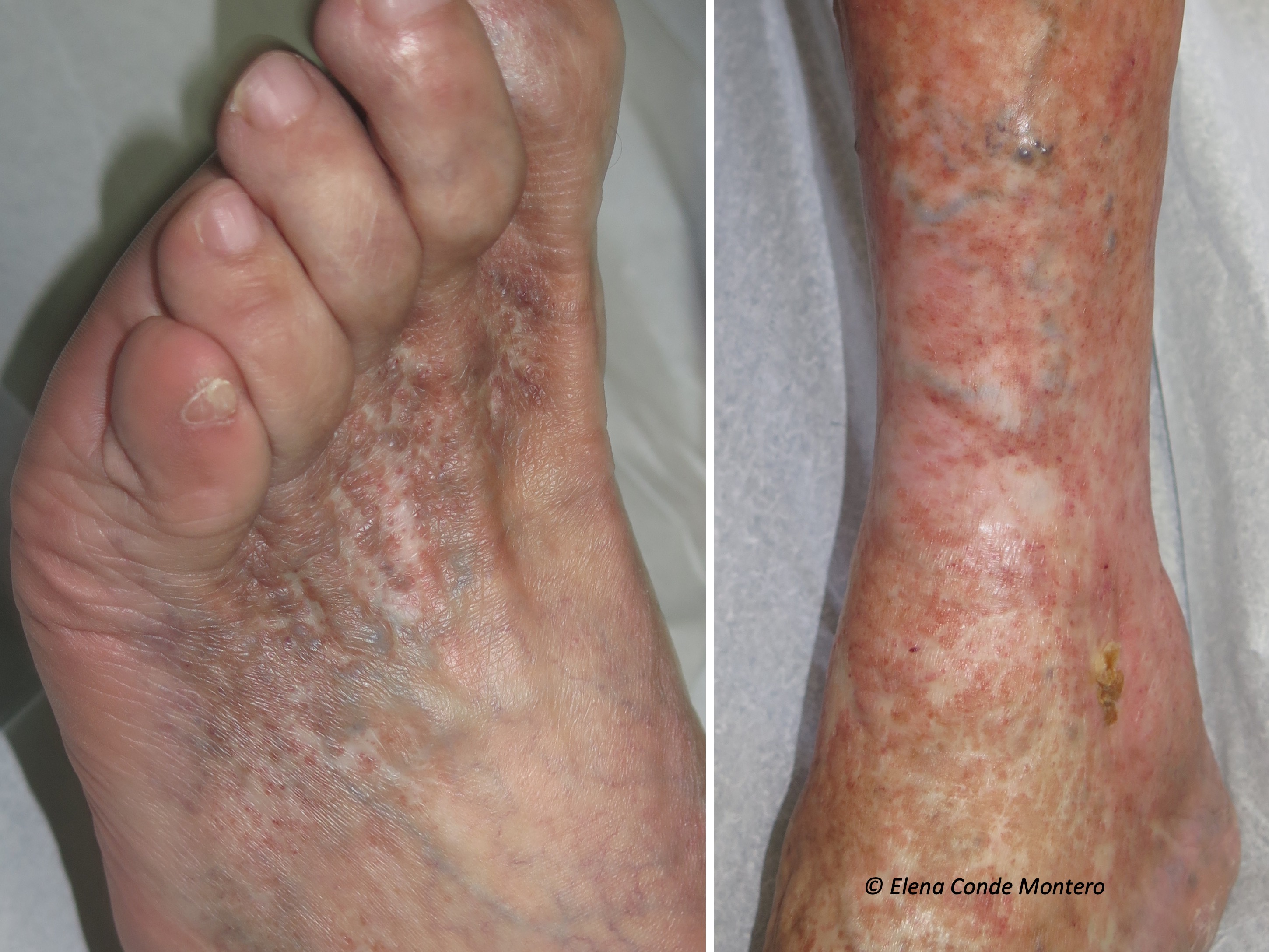
White atrophy (atrophie blanche) secondary to chronic venous insufficiency

What is white atrophy?

White atrophy (or “atrophie blanche”, which is the French term with widespread use) is the way to call atrophic stellate, porcelain white plaques, with red dots inside (dilated vessels), which can appear on the leg and foot for different reasons. That is to say, it is a term that must be used to describe a cutaneous alteration of cicatricial aspect, which is not pathognomonic of any disease, since it can appear in several clinical situations. Therefore, it is essential to make a detailed clinical history whenever we find it. White atrophy is usually painful and, if ulcers develop in that area, their healing tendency will be limited.

Why does it occur?

It is not known exactly, but it is usually associated with the alteration of the microcirculation. The authors who have histologically studied these lesions initially describe occlusions of the small vessels of superficial dermis with fibrinous microthrombi, hyalinization and wall thickening and, in lesions of longer evolution, dermal sclerosis and epidermal atrophy.

Therefore, all those diseases that produce this type of decrease in microcirculatory flow can trigger “tissue infarcts” and the consequent “scar areas”, which we call white atrophy. Chronic venous insufficiency is one of these diseases and, in fact, the one most associated with white atrophy.

However, it may appear secondarily to other disorders involving coagulation disorders (hypercoagulability states, collagen disorders, alteration of fibrinolysis) and even in a primary presentation. All of these primary and secondary causes of white atrophy would be included within the entity known as livedoid vasculopathy, a type of occlusive vasculopathy without associated vasculitis. In many patients with white atrophy and a diagnosis of livedoid vasculopathy, we also find chronic venous insufficiency. Livedoid vasculopathy usually affects middle-aged women and presents with painful purpuric papules, typically on the dorsum of the feet and inframaleolar region, progressing to ulcers and areas of white atrophy. Therefore, in cases of white atrophy without associated chronic venous insufficiency, we should perform both lab tests to exclude thrombophilia and biopsy of the edge of the ulcerated lesions.

So, why is the term white atrophy used as a synonym for livedoid vasculopathy?

Because the lesions of white atrophy are very striking in this type of occlusive vasculopathy. However, as we have seen, white atrophy is not exclusive to this disease and, in addition, livedoid pathology is an entity with many points of controversy. Conclusion, they should not be used as synonyms.

How should white atrophy be treated?

The treatment will be that of the associated disease and the objective will be, mainly, to avoid the progression of the lesions. In cases of chronic venous insufficiency, the control of venous hypertension is essential, with anti-edema measures, compressive therapy and, if possible, intravenous treatment (sclerotherapy or thermal ablation techniques). Compressive therapy must be adapted to the patient’s tolerance, as these lesions can be very painful.

Acroangiodermatitis secondary to chronic venous insufficiency

Although acroangiodermatitis (called acroangiodermatitis of Mali, the surname of the person who described it) is usually considered infrequent, this error in the estimation of its prevalence may be due to an underdiagnosed disorder. The truth is that in our wound consultation we see it frequently.

What is acroangiodermatitis?

It is a reactive proliferation of small vessels in response to a chronic vascular alteration, such as poorly controlled venous hypertension. Therefore, its most frequent location is the foot and leg. As it can also appear secondarily to arteriovenous malformations or fistulas, it can develop in upper extremities.

How does it occur?

Initially, eritemato-purpuric or brownish macules usually appear, which evolve to the formation of papules or plaques that can become warty and ulcerated. The discomfort is very variable, from asymptomatic to very painful lesions. They usually involve the back of the foot, affecting the toes and ankle, including the malleolar area. However, it can also appear on the leg. It can be uni or bilateral, depending on the evolving stage of chronic venous insufficiency in each limb.

Its clinical presentation may be similar to other diseases that should be included in differential diagnosis, especially hyperpigmentation by haemosiderin deposits or Kaposi’s sarcoma.

How is acroangiodermatitis diagnosed?

The confirmation diagnosis will be obtained by performing a skin biopsy of the lesions. Histologically, acroangiodermatitis is characterized by a proliferation of endothelial cells and an increase in the number of pericytes (perivascular spindle cells) in the dermis. The use of two immunohistochemical stains will help us to differentiate it from Kaposi’s sarcoma. CD 34 staining is only positive in endothelial cells in acroangiodermatitis, whereas in Kaposi sarcoma it also stains perivascular cells. In addition, Human Herpesvirus-8 staining is negative in acroangiodermatitis.

How is it treated?

Treatment of acroangiodermatitis secondary to chronic venous insufficiency is the control of venous hypertension. Therefore, we will insist on anti-oedema measures and the use of adequate therapeutic compression. As in the case of white atrophy, the objective will be to slow down the evolution of the disease.


References:

  1. Alavi A, Hafner J, Dutz JP, Mayer D, Sibbald RG, Criado PR, Senet P, CallenJP, Phillips TJ, Romanelli M, Kirsner RS. Atrophie blanche: is it associated with venous disease or livedoid vasculopathy? Adv Skin Wound Care. 2014 Nov;27(11):518-24; quiz 525-6.
  2. McVittie E, Holloway S. Aetiology and management of atrophie blanche in chronic venous insufficiency. Br J Community Nurs. 2015 Dec;20 Suppl 12:S8-S13.
  3. van Montfrans C, De Maeseneer MGR. Atrophie Blanche (C4b) Can Be Reversible After Targeted Treatment. Eur J Vasc Endovasc Surg. 2019 Sep;58(3):435.
  4. Badahdah HM, Edrees KM, Alnasr L, Junainah E. Acroangiodermatitis of Mali(Pseudo-Kaposi Sarcoma) Associated with Chronic Venous Insufficiency and Obesity: A Case Report. Wounds. 2018 Nov;30(11):E105-E107.