Home Blog Page 3

Why animals do not develop venous ulcers?

Surely this question has ever crossed your mind… In this post we are going to see why vets never have to treat venous ulcers, nor other signs of chronic venous insufficiency, such as stasis eczema or lipodermatosclerosis;)

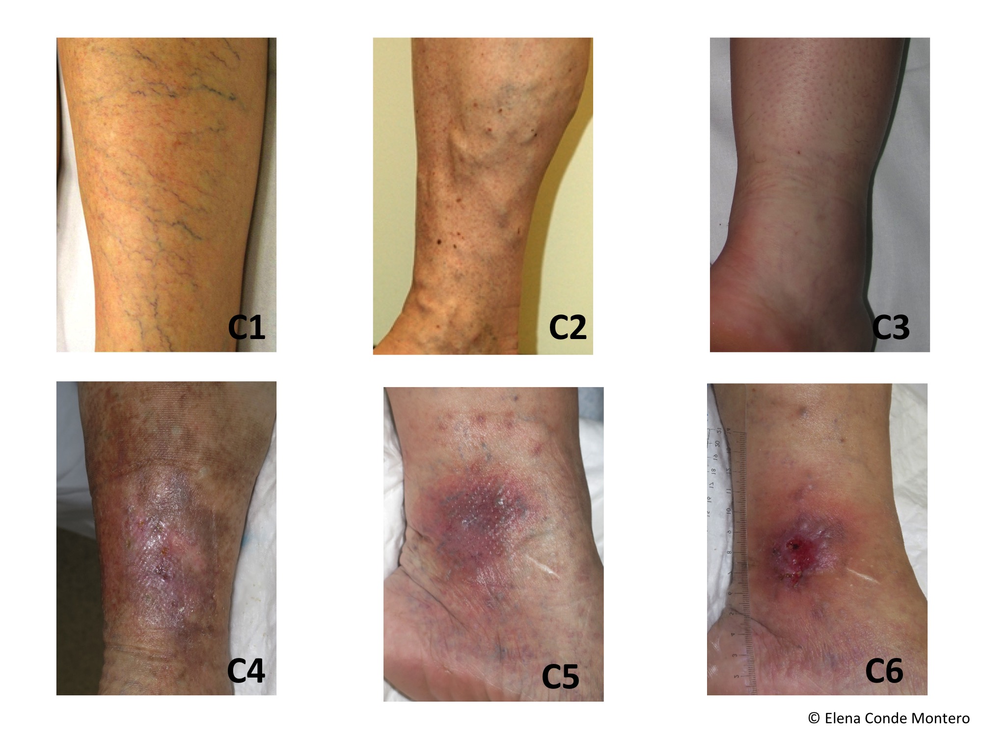
Chronic venous insufficiency (CVI) can be considered a disease unique to humans. This is mainly due to the fact that humans are bipedal, with long limbs and that our skin is elastic (we have many elastin fibres in the superficial and deep dermis). Here is a reminder of the clinical stages of the CEAP classification of chronic venous disease (which is considered CVI from C3 onwards), and I take this opportunity to recommend the post “Corona phlebectatica: sign of advanced chronic insufficiency”, where I explain the new developments in this classification;)

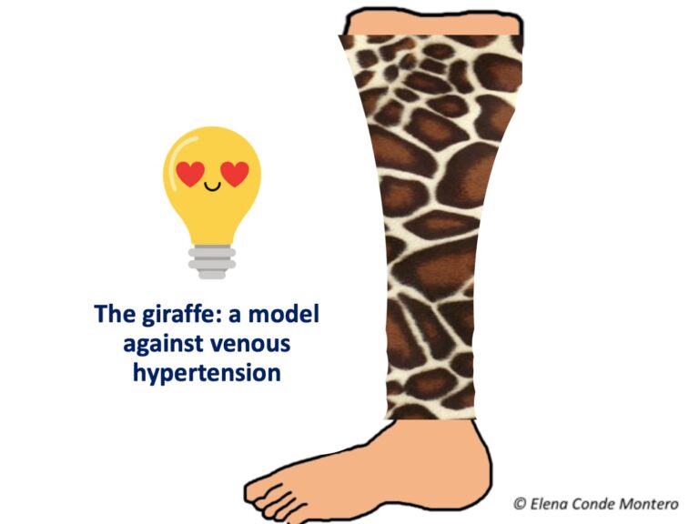
Quadrupedality is a protective factor against venous hypertension, which is the cause of venous ulcers. But this does not mean that animals are free from having varicose veins or at least distended and irregular veins… In fact, horses and cows can have them… So why don’t they develop stasis skin changes and venous ulcers? Because their leg skin is thick and has many collagen fibres and few elastin fibres (i.e. it is a tight, inelastic framework). Therefore, their skin increases the underlying tissue pressure and acts as a wall against which the muscle collides when contracting and, consequently, facilitates the valve closure of the veins, promoting venous and lymphatic return. In other words, its skin is a very efficient compression system, as it works as a bandage with a high degree of rigidity. In the post “Band and bandage: not the same thing” you will find the explanation of the elasticity of a band and the rigidity of a bandage.

The case of giraffes is very interesting. A priori, without the information I have just given, it is easy to think that, because of their height and the length of their legs, they could have chronic venous disease. However, despite having venous pressures of 250mmHg in their ankles, they do not develop varicose veins due to the inelastic characteristics of their skin. One study has shown that in the dermis of giraffe legs there is a high density of collagen fibres and little elastin (just enough to allow mobility without skin distension).1

Observation of giraffe feet inspired the design of self-adjusting compression devices with Velcro fasteners.2

So, although it is difficult to use animal models to study chronic venous disease, which is unique to humans, the identification of preventive and protective factors present in animals has clear implications for therapeutic innovation in humans.

How much we have to learn from our animal friends – Merry Christmas!

Do you know the benefits of black tea dressings?

Another post about traditional treatments, which you know is a topic I love;) For some time I had in mind to dedicate a blog post to black tea, but the lack of available evidence discouraged me from doing so… However, I have decided to do so after hearing about it in a presentation on new developments in the treatment of dermatitis at the CILAD VIRTUAL congress. Before focusing on the topic of the post, I would like to thank my friend and great wound expert, José Contreras, for proposing me to moderate with him the table on wounds at this congress, in which some of the world’s leading experts on wounds participated:)

Returning to black tea, the first thing to say is that it is a treatment that has been widely used for years among dermatologists of Germanic culture, despite the fact that, until very recently, there were no published studies on the benefit of its use. And how did I find out about this treatment if there was no evidence available?  Three years ago when I spent a stay at the University Hospital in Zurich and saw that my friend Jürg Hafner, a super wound expert, was using it.

The experience with this treatment in my clinical practice is very good. We use it mainly for erosive pustular dermatosis of the scalp and leg, where we recommend twice daily application in combination with topical corticosteroid.

Until a year ago there was nothing published in the literature on this treatment, but here is an article in which its great benefit in cases of facial eczema is evidenced.

This is a prospective, non-comparative study involving 22 patients with facial dermatitis (atopic or contact eczema). The patients applied gauzes soaked in black tea and emollient cream for 6 days. To prepare them, they infused a 2g black tea bag in 200 ml of water for 10 minutes. This first infusion was discarded and a second dilution was made with the same bag, left to cool at room temperature and the infusion was used to soak the compresses for several days of treatment. The frequency of application of the black tea was decreasing (5 times a day for the first few days, reducing to 3 applications from the third day onwards depending on the improvement). The recommended time to maintain the gauzes was 20 minutes. A highly significant reduction in all scales assessing signs and symptoms of eczema activity was found in the first three days of treatment, and this improvement continued until day 6. No adverse effects were observed.

Let’s take advantage of the references in this article to find out a little more about the properties and effects of black tea (although I’ll tell you that not much is known…).

The mechanisms by which black tea has such an interesting effect have not yet been studied, but it seems that it is not only the active principle that is interesting, but also the way it is applied. Firstly, it is known that black tea contains astringent substances (tannins) and flavonoids, with their known anti-inflammatory properties. Secondly, the application of compresses soaked in an aqueous solution reduces acute epidermal inflammation.

They can be used in combination with emollients (as in the study we have just seen) or with topical corticosteroids, which is how we use them in our practice. The frequency of application will depend on the evolution of the lesions.

An important thing to bear in mind is that black tea contains nickel. It is not clear whether its concentration is sufficient to cause hypersensitivity in patients with nickel allergy, but some German dermatologists consider it a contraindication for its use.

This inexpensive and safe treatment can be very interesting for those exudative eczemas, especially on the legs, that we find so often in our patients.


Corona phlebectatica: sign of advanced chronic venous insufficiency

The responsible for my selection of this topic is the Virtual Congress of the European Academy of Dermatology, which has been held last week. It has been a pleasure to talk about therapeutic pearls in the session on ulcers, moderated by the great expert and good friend Jürg Harner, who also talked about punch grafts. But the idea for this post did not come from the ulcers session, but from the phlebology  session (in many countries in Europe phlebology is a subspecialty of dermatology;). My dear Bibi van Montfrans talked about new developments in phlebology and among them was the recent update of the CEAP classification of chronic venous disease, with the inclusion of corona phlebectatica.

What is the corona?

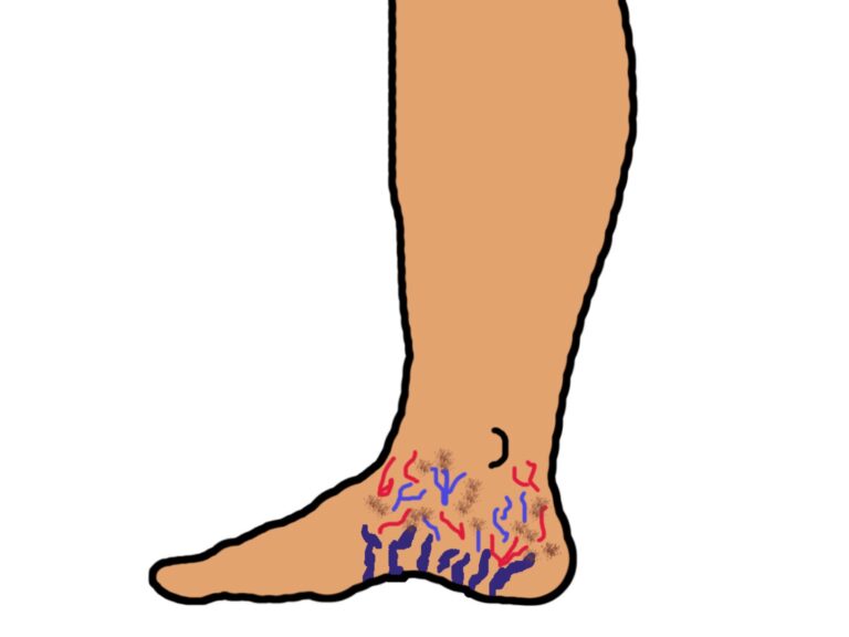
It is the group of telangiectasias, dilated veins and capillary damage (purpuric or brownish macules) secondary to venous hypertension, which are distributed in a fan pattern on the inner or outer aspect of the ankle and foot.1

It was first described in 1960 and has traditionally been considered an early sign of chronic venous disease. However, studies on this entity have led to it no longer being considered simple telangiectasias in the foot, but a clinical marker with a high predictive value for the appearance of cutaneous changes and the development of venous ulcers. In fact, corona phlebectatica is associated with the presence of saphenous reflux or incompetent perforators.1

Conclusion: We need to look at the ankles and feet of people with chronic venous disease!

What is the CEAP classification?

The CEAP (Clinical-Ethiological-Anatomical-Pathophysiological) classification was created in 1994 to describe the broad spectrum of presentation of chronic venous disease. It was modified in 2004 to refer more broadly to chronic venous disorders. In the post “CEAP classification of chronic venous disorders: let’s all speak the same language” you will find a very detailed explanation. However, that blog post is now outdated because this classification has just been reviewed and modified by international experts.2

What changes are involved in updating the CEAP classification?

One of the new features of the revised CEAP classification is the inclusion of a specific C4c section for the presence of corona phlebectatica. This recognizes that it is a prognostic marker in the evolution of chronic venous insufficiency.3

The remaining modifications are as follows:3

In case of recurrence, an r will be added at stage C2 (C2r=recurrent varices) and C6 (C6r=recurrent ulcer).

Section Es (Etiology, secondary causes) differentiates between intravenous causes, such as post-thrombotic syndrome (Esi) and extravenous causes, such as obesity (Ese).

Section A (Anatomy) has become more practical, as abbreviations for anatomical terms are now used.

I finish by talking again about my friends Bibi van Montfrans and Jürg Hafner, excellent dermatologists and better people, with whom I will participate in a Webinar organized by the European Academy of Dermatology to talk about Martorell ulcer and its star treatment 😉

References:

  1. Uhl, Jean-François & Cornu-Thénard, André & Carpentier, Patrick & Antignani, Pier. (2013). Focus on corona phlebectatica: Diagnostic, significance and predictive value in chronic venous disorders. Reviews in Vascular Medicine. 1. 38–42.
  2. Lurie F, Passman M, Meisner M, Dalsing M, Masuda E, Welch H, Bush RL, Blebea J, Carpentier PH, De Maeseneer M, Gasparis A, Labropoulos N, Marston WA, Rafetto J, Santiago F, Shortell C, Uhl JF, Urbanek T, van Rij A, Eklof B, Gloviczki P, Kistner R, Lawrence P, Moneta G, Padberg F, Perrin M, Wakefield T. The 2020 update of the CEAP classification system and reporting standards. J Vasc Surg Venous Lymphat Disord. 2020 May;8(3):342-352.
  3. Lurie F, De Maeseneer MGR. The 2020 Update of the CEAP Classification: What is New? Eur J Vasc Endovasc Surg. 2020 Jun;59(6):859-860. doi: 10.1016/j.ejvs.2020.04.020. Epub 2020 May 4.

In which situations would it be better to remove the bandage and leave the legs elevated and open air?

The title of this post reflects the best therapeutic strategy for a person with leg wounds with poor evolution due to the occlusive effect of dressings and bandages.

Most of you will ask “when does this happen and how can we identify it?” Well, the answer, as is so often the case in the field of wounds, is not to be found in guidelines, books or published studies… But these are the two situations that I encounter with some frequency in my practice: treatment-resistant erosive pustular dermatosis of the leg and excessive maceration after punch grafting.

Perhaps this is the first time you have heard the term “Erosive pustular dermatosis of the legs”. However, despite being unaware of the existence of this entity, I am sure you have seen it more than once in your daily practice.  In fact, I dedicated the following post “Pustular and erosive dermatosis of the legs: you may have seen a case without knowing it“.  Its clinical presentation does not always help in the diagnosis, as although sometimes pustules or pustular lakes may be seen, it is normal for erosions to predominate, with or without scabs. Although its aetiopathogenesis is unclear, it usually appears in older women, in the context of chronic venous insufficiency and photoageing. It has also been associated with the occlusive effect generated by dressings and bandages. This is the reason why, despite adequate treatment with topical corticosteroid (treatment of choice), zinc oxide (see post “Bandages impregnated with zinc oxide: Unna’s boot and much more” ) and compressive therapy, in some cases there is no improvement.

With regard to punch grafting, sometimes, despite adequate compression and rest with the legs elevated, in the successive dressing changes during the first weeks we do not achieve complete epithelialisation, with excessive exudate and a “congested” appearance of the grafts.

Given that the problem in both cases is occlusion, the solution is to remove the dressings and bandages and leave the legs uncovered. To control the oedema in parallel, rest with the legs elevated is essential. In cases of erosive pustular dermatosis, topical corticosteroids of moderate-high potency should be applied daily (we can also add a zinc oxide drying solution or astringent solutions) and, in the case of wounds covered with punch grafts, our experience is very good with the application of zinc oxide drying solution once or twice daily.

In a few days with this strategy we can achieve a great improvement and we can go back to compressive therapy 😉

What is the EWMA and what does it bring to my clinical practice?

Talking about the EWMA is something I have had in mind for months. I have decided right now because I want to encourage you to participate in two super interesting educational events. The first one is a Webinar about atypical wounds, scheduled for November 3rd, which we are preparing with great enthusiasm 🙂

The second is the EWMA annual flagship event (this time online, of course) on 18 and 19 November:

As is tradition, this year I will also dedicate a post to the most relevant aspects (for me, of course:) of this fantastic congress, in which I always learn a lot and collect many updated documents.

And now back to the title of the post…

What is EWMA?

The European Wound Management Association (EWMA) is a European organisation that brings together different scientific societies, groups and individuals interested in wound care, from a multidisciplinary approach.

It is a non-profit charity that was founded in 1991. It currently collaborates with more than 50 scientific societies in Europe.

Its main objective is the dissemination of good clinical practice guidelines and the support of up-to-date education of wound care professionals. In addition, EWMA works to raise awareness of the significant impact of wounds on the quality of life of individuals and the economic burden they represent for healthcare systems.

All the activities of the organisation are supervised by the EWMA Council, whose members actively participate in EWMA activities through committees and working groups. I say “we participate” because in May I was elected member of the EWMA Council 🙂  So I can tell you first hand that this Council is extremely active and committed! Kerstin Protz and Paulo Ramos have also joined this year, so we are the “new council members” :)

How does EWMA help me improve my care for people with wounds?

Essentially by providing you with different training tools:

  1. Courses on the EWMA E-learning platform

2. Documents on different aspects of the wound field, which can be downloaded free of charge. Among these documents is the one on atypical wounds, “Atypical Wounds”, led by dermatologist Kirsi Isoherranen, in which it was a pleasure to collaborate;)

3. The Journal of the European Wound Management Association. It is a journal for EWMA members that focuses specifically on EWMA activities and is published twice a year. It includes current news from the wound world, clinical experiences, peer-reviewed scientific papers and review articles. You can become a member and subscribe to the journal, but also publish articles in it! Your scientific contributions are very valuable.

4. And, of course, the Webinars and the annual EWMA Congress, with which I started the post 😉

To conclude, I would like to thank my friend Kirsi Isoherranen, for her confidence in me, support and encouragement to be part of this very enriching scientific association. Kiitos, Kirsi!!

Tricks to avoid complications in acute wounds

0

The following mental association is very common: “acute wound = wound that will close without problems”. However, an acute wound can become complicated and chronic (or even considered chronic from the moment of its onset due to comorbidities of the patient).

The essential difference between an acute and a chronic wound is that the former will heal in the expected time, while the latter presents a pro-inflammatory microenvironment that prevents the progression of the different phases of wound healing.

While for chronic ulcers there are multiple clinical guidelines, for the management of acute wounds there are no agreed protocols. In fact, the variability in the treatment of these wounds is noteworthy, especially the most frequent ones, which are abrasions, lacerations or superficial ulcers after mild trauma (which are often consulted by Whatsapp and we do not know what to recommend). Therefore, although some of the points in this post have already been covered in other entries, I think that having this “list of tricks” can be very useful to you 😉 Here we go!

  1. In clean acute wounds, we must avoid aggressive cleansing, with excessive friction. Not only is it not known whether it is better to wash with or without soap, but, as we saw in the post “The art of wound cleansing”, the  cleansing of these wounds does not seem to be associated with a lower rate of infection or other healing benefits. In fact, some experts only recommend cleansin dirty wounds (see post “Why do we continue to use topical antibiotics in clean wounds after dermatologic procedures?”)
  2. If the scab does not fluctuate and there are no signs of infection, we should not remove it. Keeping it hydrated, for example with jelly petrolatum can accelerate the healing process (See post “How do we manage clean wounds after dermatological procedures?”). The use of products with hyaluronic acid (“Reasons for the hyaluronic acid boom in wound healing”) or zinc (“Why do we use topical zinc on wounds and perilesional skin?”) can also help accelerate epithelialisation.
  3. Superficial burns are characterised by excessive inflammation. This prolonged inflammatory phase not only delays the healing process but also increases the risk of hypertrophic scarring. Although more studies are needed, topical corticosteroids is a very interesting treatment alternative (See post: “Does it make sense to apply topical corticosteroids to improve the healing of superficial burns?”)

4. In clean acute wounds the use of topical antiseptics and antibiotics is of no interest (See post “Why do we continue to use topical antibiotics in clean wounds after dermatologic procedures?”)

5. In superficial exuding wounds, zinc sulphate foments 1:1000, once or twice a day, are of interest because of their astringent power (see post: “Gauzes soaked with astringent and antiseptic foments for wounds and perilesional skin”)

6. No dressing has shown superiority over the others (in fact bioactive dressings are mainly used, in the clinic and in research studies, in chronic ulcers). However, in bleeding wounds, calcium alginate is very beneficial because of its haemostatic power and its ability to “simulate a physiological crust” (See post: “Why do we use so many alginate fibre sheets in our wound clinic?”).

7. If coverage with dressings is required, dressing changes should be spaced as far apart as possible (depending on exudate and discomfort control). We must remember that at each dressing change we remove the excess exudate and denatured tissue, but we also remove growth factors and cells that promote healing).

8. As long as there are no contraindications (see post “New recommendations about compression therapy”), any acute leg wound will benefit from compressive therapy, because of its anti-gravity and anti-inflammatory effect, even if the person does not suffer from chronic venous insufficiency. (See postCompression therapy after dermatologic surgery on the leg”). We will also recommend anti-oedema measures, such as rest with elevation of the lower limbs.

9. We will avoid the tension forces that separate the edges of the wound, since they make it difficult to contract the wound and promote the appearance of a pathological scar (see post “The myofibroblast: the great ignored in wound healing”).

10. Finally, remember that the evolution of a wound will always depend on the comorbidities of the patient (smoking, age, immobility, nutritional status, cognitive degeneratio, dermatoporosis, chronic venous insufficiency, diabetes…). Therefore, there are wounds that, despite starting out as minor injuries, after trauma that are sometimes so minor that they are not even remembered, should already be considered as ulcers that are going to have a torpid evolution (See posts: “Wounds secondary to dermatoporosis or chronic skin insufficiency”, “Large leg ulcers after mild trauma “).


How to close large ulcers after mild trauma in the elderly?

How many times have we asked ourselves this question, health professionals, caregivers and, of course, the people who suffer them on their own skin?

Without doubt, there are many ways of doing things (especially in the field of wound healing) and in most cases we don´t know which is the best alternative. However, in this type of ulcer, in older patients, with multiple comorbidities, the most frequent management is conservative, with the use of products that fail to promote healing and with the consequent associated complications (poor pain control, infections, hospitalization). The impact of these ulcers on the quality of life of elderly people is enormous. In many cases we find people over 90 years old, without any associated disease, to whom the onset of such a wound, after a mild traum, changes their lives. They stop walking, isolate themselves, become depressed, suffer unbearable pain that has to be treated with painkillers with many side effects… 

As we commented in the post “Large leg ulcers after mild trauma”, age, hypertension and diabetes favour the process called arteriolosclerosis, which consists of the thickening and decrease in elasticity of the walls of the small arteries (arterioles), in some cases with the presence of calcifications. As I also explained in that post, arteriolosclerosis could be the trigger for the vicious necrosis-inflammation circle, which causes these large ulcers after minimal trauma.

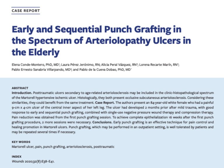
Taking into account that we consider that Martorell ulcer and post-traumatic arteriolopathy associated to age share clinical aspects and both present arteriolosclerosis, we could consider both in the same disese spectrum that benefit from the same treatment, that is, punch grafting ?See post “Large leg ulcers after mild trauma”, Interest of early punch grafting in Martorell hypertensive ischemic ulcer”

We always try to prepare the wound bed as well as possible to increase the percentage of graft taking. However, pain, often associated with a certain degree of cognitive impairment, makes adequate debridement difficult. Our solutions to this frequent problem are irrigation with sevoflurane, as an analgesic and vasodilator (“Have you ever heard of topical sevoflurane in wounds”) and early and sequential punch grafting in suboptimal beds. Sequential punch grafting (every 2-3 weeks) implies that with the first sessions we can achieve a progressive limitation of necrosis, wound contraction, decrease of pain and increase of granulation tissue, which will favour the attachment of subsequent grafts. That is, even if no graft gets attached on an initial bed with a lot of slough, this early coverage will promote the release of cells and growth factors essential for healing, in addition to having a significant analgesic effect (See post “Punch grafting as a painkiller for painful ulcers”)

Other allies in this anti-inflammatory and pro-epithelialisation strategy for these post-traumatic ulcers due to age-related arteriolopathy (which we also use for the Martorell ulcer) are:

I finish this post by sharing a case we have just published, which I hope you will find inspiring to help your patients 🙂

early and sequential punch grafting arteriolosclerosis

What type of compression therapy to choose in each situation?

Once again, the post in summer time has compression therapy as its main theme. Last year we talked about the “Essential Decalogue in the fight against venous hypertension”https://elenaconde.com/en/essential-decalogue-in-the-fight-against-venous-hypertension/. This time we will also talk about compression in a very practical way. Here you may find attached an article that we have just published, with theoretical but undoubtedly practical aspects of compression therapy.
S0001731020301903

In this publication we describe the characteristics of the different compression therapy devices that we commonly use in the practice, both in the prevention and treatment of venous ulcers. It is always a good time to remember that compressive therapy is the best antihypertensive and anti-inflammatory treatment for any leg ulcer as long as there are no contraindications.

Although I have dedicated some posts in a monographic way to some of these devices (“Zinc impregnated bandages: Unna boot and much more“, “Why are adjustable compression wraps so popular?”) I think it is very useful to have a little scheme of “when to select one or the other, that is, in what kind of patients or situations is more beneficial some of them… So here you have it:)

Before going to the schematic, remember that all systems can be used with or without an ulcer. However, in large ulcers, we will normally recommend bandaging. In the case of small ulcers, which require frequent dressing changes, stockings or the adjustable compression wraps are good alternatives.

With this post I say goodbye until September with this beautiful watercolor that a very special patient has given us… If there is a word that for me defines perfectly our wound clinic is “EQUIPAZO”… How nice when we are also seen like this by people we love to help 🙂

Povidone-iodine and chronic ulcers: a great controversy

Povidone-iodine is a product that is used by many professionals who treat wounds, but it also has many detractors… Despite not being new and having an enormously extended use, there is much ignorance and debate about this antiseptic. Surely you have asked yourself some of these questions about povidone-iodine: Why is it so widely used in clinical practice? What type of wound and person is it for? What differences are there between its different presentations? Is it really as cytotoxic as people think?

In this post we will review and analyse the available evidence on the usefulness and safety of using povidone-iodine in chronic wounds. A group of Asian experts with extensive experience in wound healing met in 2016 to evaluate the available published evidence on povidone-iodine and to share their experience with this cheap and widely used product in order to reach a consensus of recommendations in Asia.1 Therefore, much of the information in this entry is taken from that interesting document.

What is povidone-iodine?

Povidone-iodine is a chemical complex composed of povidone (water-soluble polymer), hydrogen iodide and elemental iodine. It is therefore an iodophor, i.e. an iodine carrier.

Despite the fact that the history of povidone-iodine is little more than 60 years old, since the beginning of the 19th century the use of iodine was quite widespread in the prevention and treatment of skin infections and wounds, due to its recognized broad-spectrum microbicidal action. However, iodine compounds existing until then, such as tincture of iodine, produced irritation, cellular toxicity and high skin pigmentation. With povidone-iodine these adverse events largely disappear, as this complex provides a reservoir for the continuous release of small amounts of free iodine. This free iodine, slowly released from the povidone-iodine complex, kills eukaryotic or prokaryotic cells by altering the cell membrane and penetrating the cytoplasm by denaturing proteins, fatty acids and nucleotides. As it is a slow release, the concentration of free iodine is low, so its toxicity to human cells would be minimized.1,2

What has been studied about its microbicidal activity, its penetration into biofilms and its generation of resistance?

Its microbicidal efficacy has been studied in vitro. It has demonstrated a wide spectrum of activity against Gram+ (including MRSA), Gram-, fungi, viruses and protozoa.1

Regarding its action on biofilms, a recent in vitro study has shown its efficacy, superior to other antimicrobials, in eradicating biofilms of Pseudomonas aeruginosa, MRSA and Candida albicans.3 But I repeat that these studies have been conducted in vitro, so they may not be reproducible in vivo. In fact, certain proteins present in wound exudate may inactivate iodine and thus limit its microbicidal effect, something that requires further evaluation in new in vivo studies.

On the other hand, discussing the bacterial resistance that povidone-iodine might generate is essential, as the large increase in resistance to antibiotics (and different antiseptics) is one of the major clinical and public health problems worldwide. However, despite its widespread use, the development of povidone-iodine resistance has not been described.1

How does the presentation of povidone-iodine influence its action?

Povidone-iodine has different presentations. The rate of iodine release and the changes that will occur in the wound microenvironment depend on the composition of the excipients (a drying solution is not the same as a moisturizing gel or ointment). The concentration and formulation of the active ingredient are important factors that contribute to the effectiveness of any antiseptic. The bactericidal component of povidone-iodine is free iodine, the levels of which are dependent on the concentration of the solution. The concentration of free iodine increases with increasing dilutions of povidone-iodine, i.e. the dilution weakens the binding of iodine to the carrier, resulting in an increase of free iodine in the solution. This would explain the paradox of the rapid antibacterial action of povidone-iodine with increasing dilution.1 The concentration required for different wound types and the recommended and necessary contact time with the tissue remain controversial. 

Are allergies to povidone-iodine common?

There is much debate among dermatologists about the allergenic and irritant potential of povidone-iodine. Actually all antiseptics can irritate the skin if used improperly, particularly on skin with eczema, in occlusion and at high concentrations. Povidone-iodine contact dermatitis has never been considered a frequent adverse event, especially in view of its widespread use. Isolated descriptions of contact skin reactions, both irritative and allergic, have been published. However, as there is a lack of consensus on the concentration and vehicle of iodine compounds to be used for diagnostic epicutaneous testing, it may be difficult to discriminate between the two types of contact dermatitis (allergic-irritative).4

What do the studies say about its influence on healing, and is it really as cytotoxic as thought?

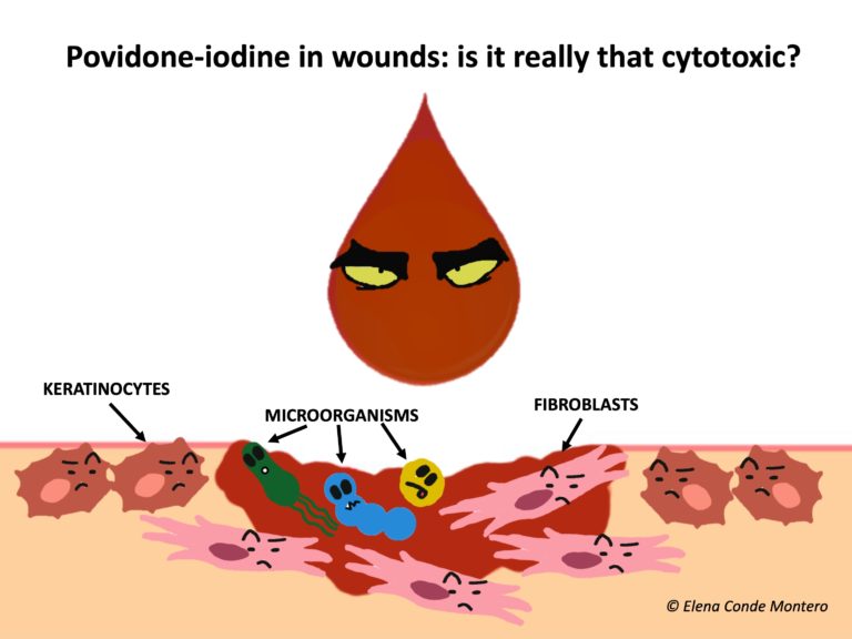
The effect of povidone-iodine on healing has been widely discussed. Studies with povidone-iodine in vitro and in animals (mainly rats) have shown variable, some conflicting results, and most have been conducted for more than 20 years.1 In vitro studies demonstrate dose-dependent cytotoxicity of povidone-iodine in cell cultures, including keratinocytes and fibroblasts.5,6 However, in mice it has been found that its use produced a faster and more complete neovascularization than other antiseptics, with a positive effect on healing.7 In fact, it has been found that concentrations equal to or less than 10% (the most commercialized in our environment), do not inhibit the processes of granulation and epithelization.1

It should be noted that experimental conditions also have a clear impact on the results. For example, the additives added to the medium may themselves promote toxicity. In fact, the pronounced cytotoxicity demonstrated in certain in vitro studies has not been confirmed in human studies, where it has been found to be safe at adequate concentrations. Human studies are scarce, with few patients included and many limitations. The different published studies include burns, pressure ulcers, venous ulcers, trauma, lacerations, skin grafts. Those studies comparing povidone-iodine with other dressings showed no inferiority in healing rates and some even found superiority. Single studies that have studied its antimicrobial activity in chronic ulcers have found decreased bacterial growth.1,8

Conclusion: We need more well-designed human experimental studies to better understand the disparities between in vitro and animal studies.

And the toxicity from its systemic absorption?

It is not clear how much povidone-iodine and how often it is applied that can develop systemic toxicity. Caution is essential in patients with thyroid disease, pregnant women and children.1,2

In what situations do I use povidone-iodine in chronic wounds?

In those situations where the highest priority is to avoid bacterial growth and therefore also to promote an astringent environment (the excipient povidone-iodine solution has a drying action). The most frequent situation would be arterial trophic lesions pending revascularization or amputation, or those who have no option of any of these surgical alternatives due to the poor general condition or complex situation of the patient.

Another interesting use of povidone-iodine solution is its application in heel pressure ulcers with the presence of dry necrotic scab, adhered to the wound bed, without fluctuation. In these cases, if the ulcer is superficial and vascularization is adequate, this scab could promote underlying epithelialization. On the contrary, if the ulcer is deep and extensive, there is associated arteriopathy and/or the patient’s complexity situation does not allow another attitude that favors healing, the objective will be exclusively to maintain the protective scab to avoid superinfection. Moreover, regarding pressure ulcers, povidone-iodine is also of interest to people in a situation of great complexity or end of life, with high risk of infection and need for frequent dressing changes.

Finally, this priority of antiseptic and drying action can also be found in infections of chronic wounds with systemic impact, with drainage of purulent material, where frequent dressing changes with thorough cleansing and application of antiseptic will be excellent adjuvants to the systemic antibiotic to control the infection.

What is your experience with povidone-iodine?

References:

  1. Bigliardi P, Langer S, Cruz JJ, Kim SW, Nair H, Srisawasdi G. An Asian Perspective on Povidone Iodine in Wound Healing. Dermatology. 2017;233(2-3):223‐233.
  2. Sibbald RG, Leaoer DJ, Queen D. Iodine Made Easy. Wounds International 2011; 2(2): Available from http://www.woundsinternational.com
  3. Hoekstra MJ, Westgate SJ, Mueller S: Povidone-iodine ointment demonstrates in vitro efficacy against biofilm formation. Int Wound J 2017;14:172–179.
  4. de la Cuadra-Oyanguren J, Zaragozá-Ninet V, Sierra-Talamantes C, Alegre de Miquel V. Postsurgical contact dermatitis due to povidone iodine: a diagnostic dilemma. Actas Dermosifiliogr. 2014;105(3):300‐304.
  5. Kashiwazaki J, Nakamura K, Hara Y, Harada R, Wada I, Kanemitsu K. Evaluation of the Cytotoxicity of Various Hand Disinfectants and Ozonated Water to Human Keratinocytes in a Cultured Epidermal Model. Adv Skin Wound Care. 2020;33(6):313‐318.
  6. Liu JX, Werner JA, Buza JA 3rd, Kirsch T, Zuckerman JD, Virk MS: Povidone-iodine solutions inhibit cell migration and survival of osteoblasts, fibroblasts, and myoblasts. Spine (Phila Pa 1976) 2017, Lachapelle JM, et al. Antiseptics in the era of bacterial resistance: a focus on povidone iodine. Clin Pract. 2013;10(5):579-92.
  7. Langer S, Botteck NM, Bosse B, Reimer K, Vogt PM, Steinau HU, et al: Effect of polyvinylpyrrolidone-iodine liposomal hydrogel on wound microcirculation in SKH1-hr hairless mice. Eur Surg Res 2006;38:27–34.
  8. Bigliardi PL, et al. Povidone iodine in wound healing: A review of current concepts and practices. Int J Surg. 2017;44:260-8.


New recommendations about compression therapy

There’s a big news flash! A very relevant consensus document on compression therapy has just been published: Risks and contraindications of medical compression treatment – A critical reappraisal. An international consensus statement.

This is a critical review, up to November 2017, including 62 publications on adverse events associated with compression. With the data obtained, 15 SUPERexperts have agreed on 21 recommendations regarding contraindications to compression therapy and strategies to reduce the risk of adverse events.

Before moving to the recommendations, I would like to emphasize that, in this review, the adverse events most frequently associated with compression therapy are skin irritation, discomfort and pain. Exceptionally, severe injuries, such as nerve damage or soft tissue involvement, such as necrosis, have been reported.

How to prevent adverse events secondary to compression therapy?

Recommendation 1: “We recommend that every patient receiving compression therapy should be screened for conditions that increase the risk of complications, and every compression device should be checked for appropriate fit and application. Contraindications for compression treatment must be considered to limit the risk of side effects”.

How to control skin irritation and “marks” associated with compressive therapy?

Recommendation 2: “We recommend using adequate skin care to prevent skin irritation in patients with sensitive skin”.

The control of stasis eczema with corticoids and zinc oxide, use of emollients, adequate padding and the use of protective skin tissues directly on the skin under the compression devices are our best strategy in the clinical practice.

Although allergic reactions to materials used in compression devices are rare, how can they be avoided?

Recommendation 3: “To prevent allergic skin reactions due to compression devices, we suggest avoiding potentially allergenic substances and dyes in compression materials”.

How do you make compression therapy comfortable for the patient?

Recommendation 4: “In patients with discomfort and/or pain below compression garments, we recommend checking the correct indication, pressure level, material, fitting or bandage techniques as well as the correct donning and doffing”.

How to control the edema in the forefoot and toes?

Recommendation 5:In patients with, or in those developing, forefoot or toe oedema when wearing compression, we suggest considering forefoot and toe bandaging or forefoot and toe compression pieces in addition to leg compression with a foot piece”.

Can I use compression in cases of local wound infection? What if the infection is systemic?

Recommendation 6: “In patients with bacterial or fungal infection beneath the compression device, we recommend considering treatment with topical antiseptics or topical antimicrobiological medication. In patients with systemic symptoms (fever, chills), erysipelas or cellulitis, we recommend that systemic treatment should be given. In other cases of systemic symptoms and severe local wound and tissue infection, the decision on the further treatment, including also medical compression, should be individualised on the basis of the local and general patient condition evaluation”.

Recommendation 7: “If the compression application or material is suspected to contribute to the infection (e.g. lateral pressure on toes with interdigital maceration), we suggest a modification of compression”.

For example, medical compression stocking with open toe or adjustable velcro devices.

How to avoid pressure injuries in risk areas?

Recommendation 8: “We suggest considering that, according to the Law of Laplace, the local pressure below the compression material may be higher than expected at bony and tendinous prominences such as above ankles, the tibia, the fibular head or above tendons such as the Achilles tendon, and to check those locations for skin lesions due to pressure”.

Recommendation 9: “To prevent tissue damage or necrosis and nerve damage in regions with a small radius, we suggest protecting these regions (tendons, nerves and bones) from inappropriate high pressure, particularly in patients with sensitive skin, by:

  • Decreasing the local pressure by inserting soft padding material
  • Using low overall pressure
  • Taking appropriate circumference measurements so that the compression devices fit properly”.

Recommendation 10: “We suggest specific precaution (padding, special care of fit, low pressure), and close controls at the initial stages of compression therapy in patients with polyneuropathy and elderly patients with frail, atrophic skin (dermatoporosis)”.
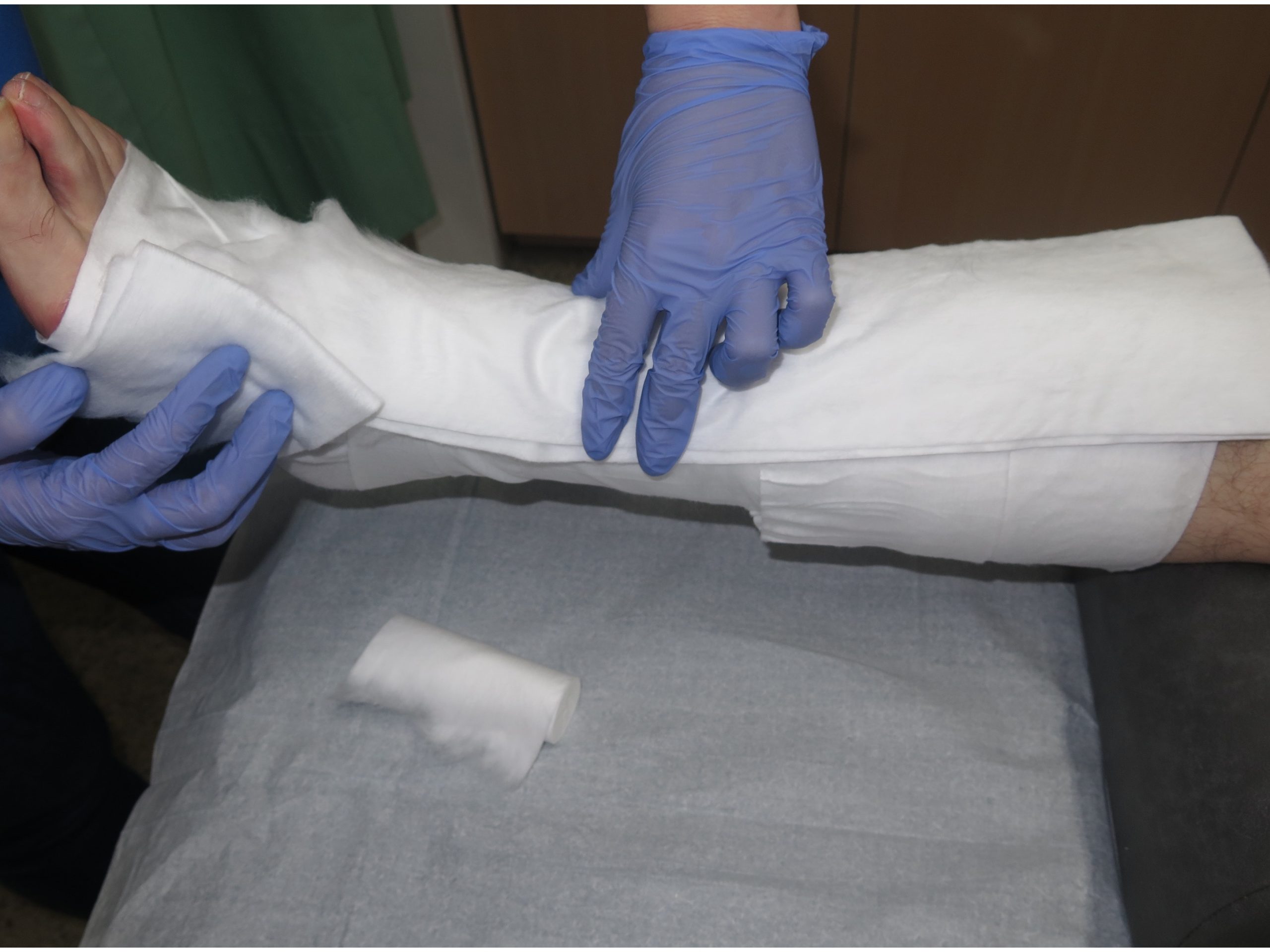
How do you avoid causing nerve damage?

Recommendation 11: “We suggest considering that pressure-induced nerve damage may occur at specific points of the leg (e.g. fibular head) mainly in cases with excessive local compression pressure, e.g. due to ill-fitting medical compression stockings, thromboprophylactic stockings or compression bandages. Numbness and nerve palsy may occur. We suggest preventing high or continuous local pressure in regions with a risk of nerve compression as well as correct sizing and application of compression. Patients at higher risk for nerve damage (e.g. patients with diabetes, patients with neuropathy) should be treated with special caution to prevent nerve damage”.

When and how to apply compression therapy in a patient with artery disease?

Recommendation 12: “We recommend checking the arterial circulation status before any kind of compression therapy is initiated. If foot pulse and/or ankle pulse is weak or not palpable, ABPI should be measured and calculated prior to initiating medical compression therapy”.

Recommendation 13: “Severe PAOD (systolic ankle pressure <60 mmHg, toe pressure <30 mmHg) is a contraindication against compression therapy with medical compression stockings. In compression bandages, the applied pressure and the elasticity of the material are important. This contraindication does not apply to intermitent pneumatic compression and to patients with non-critical leg ischaemia treated with inelastic material applied with low resting pressure”.

Recommendation 14: “In every patient with impaired perfusion of the lower limb (ABI <0.9), the clinical effect of the medical compression stockings on leg blood supply should be carefully monitored. If the situation is not recognised, there is a possibility of developing non-healing skin breaks even under low pressure medical compression stockings”.

Can we apply compression therapy after bypass surgery?

Recommendation 15: “After bypass surgery with improved peripheral arterial pressures, medical compression treatment may be performed if there is no direct compression effect on the bypass itself. We suggest avoiding the compression of epifascial bypass conduits. As for all patients with chronic leg ischaemia, the recommendations regarding the use of medical compression treatment should be followed (see recommendations 12–14)”.

Is there a risk of venous thrombosis from poorly applied compression therapy?

Recommendation 16: “Because of a tourniquet effect, improper compression can cause local superficial venous thrombosis, especially in combination with prolonged sitting (long-haul flights). To prevent thromboembolic complications, we recommend avoiding a tourniquet effect and strangulation by inappropriate application of medical compression stockings, thromboprophylactic  stockings and bandages”.

In case of cardiac insufficiency, what should I look out for?

Recommendation 17: “We recommend against applying compression in severe cases of cardiac insufficiency (NYHA IV). We also suggest against routine application of MCS in NYHA III cases. When needed, careful use of compression therapy in this patient group may be considered if there is a strict indication, with clinical and haemodynamic monitoring. In less severe cases, cautious increase of compression pressure only leads to very short phases of increased cardiac load and may lead to a substantial reduction of peripheral oedema”.

Is it appropriate to include compression in the treatment of superficial or deep vein thrombosis?

Recommendation 18: “We recommend considering that, in contrast to previous concepts, compression is not contraindicated in acute thrombotic events, but results in favourable clinical outcomes when applied with caution. In the hands of experts, proper compression leads to an immediate improvement of pain and oedema”.

What are the benefits of compression in inflammatory diseases, such as vasculitis, or infectious diseases, such as cellulitis?

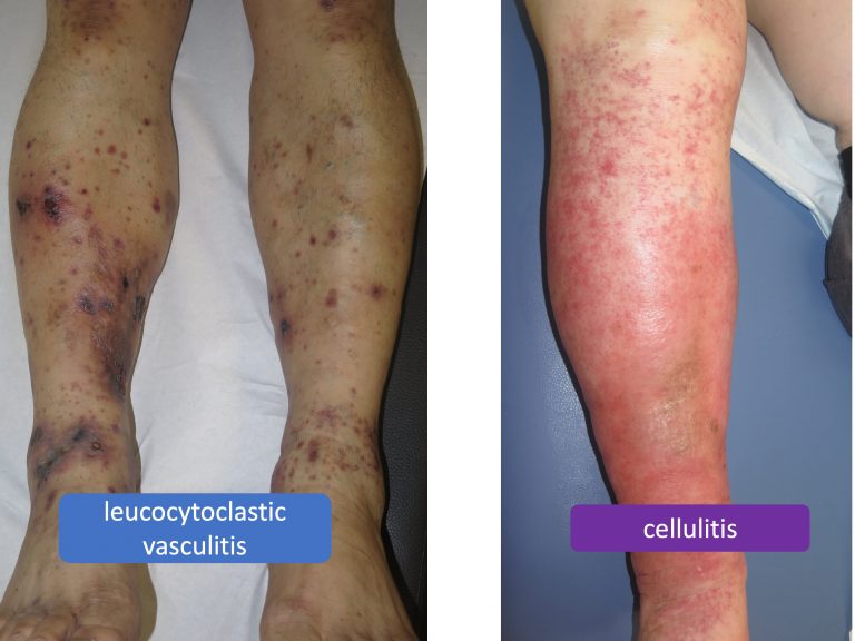
Recommendation 19: “We suggest additional compression, in purpura due to leucocytoclastic vasculitis and in leg erysipelas or cellulitis, to reduce inflammation, pain and oedema. In infectious inflammation, we suggest compression only in combination with antibacterial treatment.”
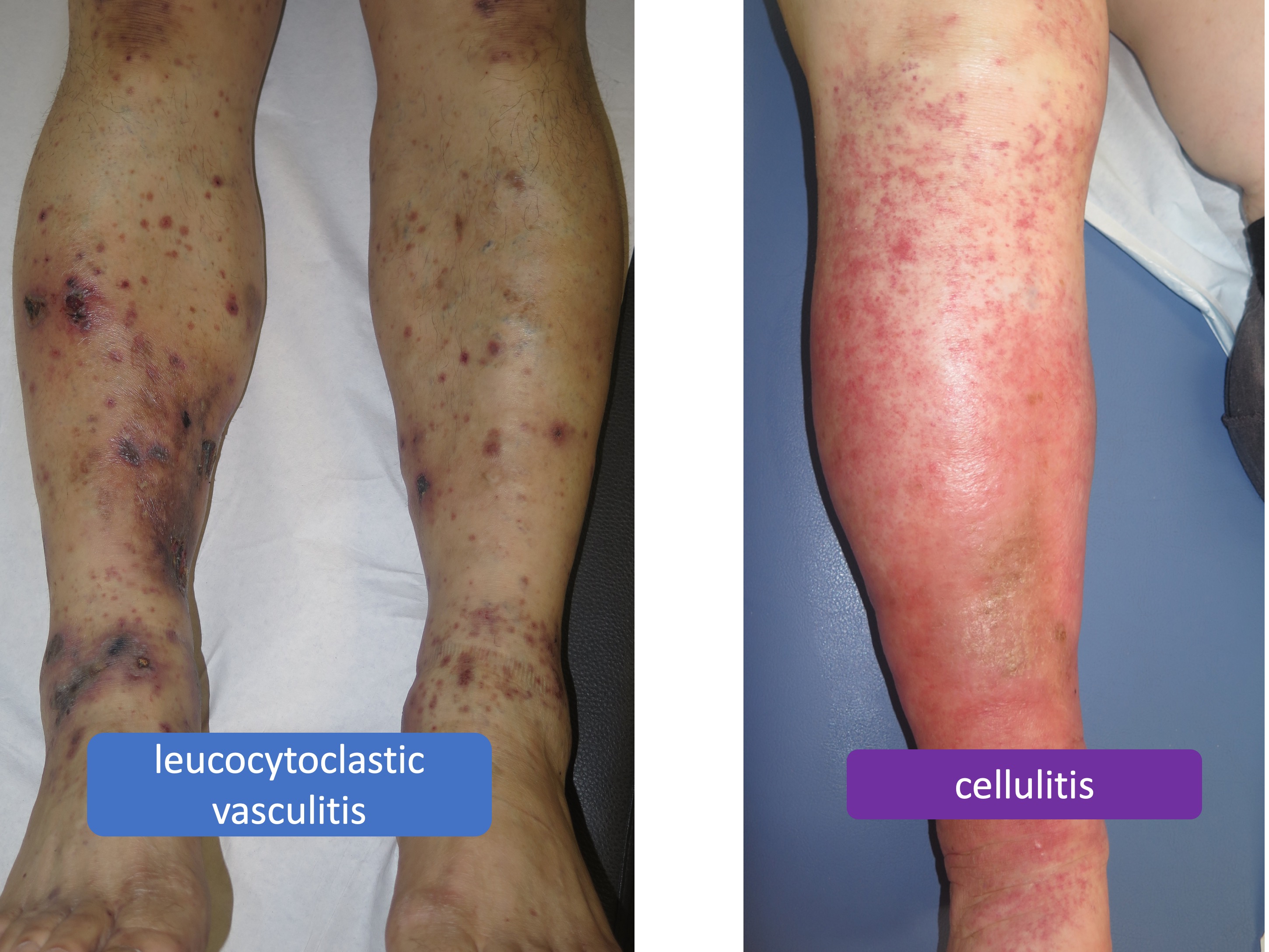
Recommendation 20: “Special precautions have to be taken if medical compression treatment is considered in patients with ‘borderline indications’. Treatment decisions should be taken on a case-by-case basis and under consideration of a careful benefit–risk assessment. In case of a favourable assessment, we suggest the use of low-pressure compression, the use of modified-compression strategies (compression materials) and the use of padding to reduce pressure peaks”.

So, for this group of SUPEREXPERTS what would be the absolute contraindications of compressive therapy?

Recommendation 21: “We recommend considering the following contraindications for sustained compression with thromboprophylactic stockings, adjustable compression wraps, medical compression stockings and elastic compression bandages:

  • In patients with severe PAOD with any of the following: ABPI <0.6; ankle pressure <60 mmHg; toe pressure <30 mmHg; transcutaneous oxygen pressure < 20 mmHg
  • Suspected compression of an existing epifascial arte- rial bypass
  • Severe cardiac insufficiency (NYHA IV)
  • Routine application of MC in NYHA III without strict indication, and clinical and haemodynamic monitoring
  • Confirmed allergy to compression material
  • Severe diabetic neuropathy with sensory loss or micro- angiopathy with the risk of skin necrosis (this may not apply to inelastic compression exerting low levels of sus- tained compression pressure (modified compression))”.

This means severe PAOD, compression of epifascial arterial bypasses, severe cardiac insufficiency and true allergy to compression material.