Home Blog Page 2

Little is said about arteriolosclerotic ulcers

I think the title I have chosen sums up perfectly the situation of this type of wound: it is much more frequent ulcer than we think, but it is underdiagnosed and we do not usually call it by its name.

As the EWMA has just organized a Masterclass in which I have talked about this topic, it is the perfect time to dedicate a post to these ulcers secondary to small vessel disease.


What is cutaneous arteriolosclerosis?

It is defined as a thickening of the wall of the arterioles of the deep dermis and subcutaneous cellular tissue. The term “cutaneous arteriolosclerosis” includes different entities in the literature: proliferative and hyaline arteriolosclerosis, which is ubiquitously found in untreated hypertension, and fibrosing endarteritis and medial calcinosis of Mönckeberg, linked to aging. Therefore, arteriolosclerosis can be found in legs of any elderly or hypertensive person, it is not specific to patients with wounds.1

What is the clinical presentation of arteriolosclerotic ulcers?

These are very painful leg ulcers, with signs of necrosis, erythematous-violaceous borders, which can progress rapidly. Since this description could fit perfectly with arterial ulcers, the first thing to do when exploring an ulcer with these characteristics is pulse palpation. People with arteriolosclerotic ulcers will always have palpable pulses.

What is the difference between arteriolosclerotic ulcer and Martorell ulcer?

There is no difference. In fact, when talking about arteriolosclerotic ulcer, the first thing that comes to mind is Martorell ulcer. The so-called hypertensive ischemic Martorell ulcer is a lesion secondary to subcutaneous arteriolosclerosis (see post: “Key elements to understanding Martorell ulcer”).

But in the clinical-histopathological spectrum of Martorell ulcer, and therefore of ulcers due to arteriolosclerosis, there is also, on the one hand, calciphylaxis, as we commented in the post “Martorell ulcer and vitamin K antagonists: a dangerous combination” and, on the other hand, ulcers due to age-associated arteriolopathy (See post “Large leg wounds after mild trauma”). In fact, while the history of chronic renal insufficiency is a differentiating factor for the diagnosis of calciphylaxis, there is no particularly distinguishing feature between ulcers due to age-related arteriolopathy and the ulcer known as Martorell ulcer.

There are fewer and fewer deaths due to major cardiovascular events, so we are more frequently encountering minor cardiovascular events, such as ulcers due to arteriolosclerosis. In fact, with the increased control of diabetes and hypertension, the clinical and histologic presentation of arteriolosclerosis ulcers in most cases is not as striking as in the early descriptions of Martorell ulcer in the literature.

We will perform a biopsy of the edge of the lesion in case we want to rule out other etiologies (See post: “Necrosis and violaceous edges in leg ulcers: keys to guide its diagnosis“).

What is the treatment?

The management of ulcers due to arteriolosclerosis is the same as that discussed in other posts on Martorell ulcer and ulcers due to arteriolopathy associated with age (See post: “How to close large ulcers after minor trauma in the elderly”).

In addition to the control of risk factors such as diabetes mellitus or hypertension, it is essential to control pain, stop necrosis and promote epithelialization. For this, in our practice we use sequential punch grafting, with or without negative pressure therapy, with excellent response.2 Sevoflurane irrigation is another alternative strategy that can help control pain and promote vasodilatation in the wound bed. (See post: “Have you ever heard of the use of topical sevoflurane in wounds?“)

I recommend you the posts: “How to close large ulcers after mild trauma in the elderly?” , “Interest of early punch grafting in Martorell hypertensive ischemic ulcer” and “Punch grafting as a painkiller for painful ulcers”.

Can compressive therapy be used?

Yes, of course! Always adapted to the needs of each person

In these patients, as the pain is extreme and constant, there is a decrease in calf muscle pumping due to a significant reduction in gait (patients stop walking to avoid pain), leading to edema and decreased skin perfusion. This edema may also explain why pain increases in these patients when standing. Therefore, compression will increase perfusion by reducing edema and improve pain. In addition, its use will enhance graft attachment (see post “Compression is key to treating leg wounds”).

Ideally, compression devices with a high stiffness index (short stretch bandages or multicomponent bandages) should be used to obtain a low resting pressure, and pressure peaks during walking. The level of compression depends on the patient’s tolerance, but 20-30 mmHg may be sufficient (see post: “What type of compression therapy to choose in each situation“).

References:

  1. Monfort JB, Cury K, Moguelet P, Chasset F, Bachmeyer C, Francès C, Barbaud A, Senet P. Cutaneous Arteriolosclerosis Is Not Specific to Ischemic Hypertensive Leg Ulcers. Dermatology. 2018;234(5-6):194-197. 
  2. Conde-Montero E, Pérez Jerónimo L, Peral Vázquez A, Recarte Marín L, Sanabria Villarpando PE, de la Cueva Dobao P. Early and Sequential Punch Grafting in the Spectrum of Arteriolopathy Ulcers in the Elderly. Wounds. 2020 Aug;32(8):E38-E41. 


Should wounds ALWAYS be cleansed

The answer is NO. As many of you will have been surprised by this answer, I think this topic deserves a post:)

You have probably already read phrases like this in some of the posts dedicated to punch grafting: “it is better to space dressing changes as much as possible and, at each dressing change, cleanse and touch as little as possible”. We even reposition the grafts that have remained in the dressing… (See post: “Repositioning and not removing the scabs: two unwritten rules of punch grafting”).

Let’s understand the reason for this recommendation.

Each dressing change is an opportunity to promote, with cleansing and debridement, the removal of what is hindering healing (excess exudate with proinflammatory cytokines, non-viable tissue, bacterial load). However, any manipulation of the wound bed will also have an impact on the cells and growth factors that are promoting wound closure.

Wound exudate, especially in acute wounds and those that are doing well, is a source of cells and growth factors that promote healing. Therefore, in these cases, frequent and/or aggressive washing with friction should be avoided, as we can interrupt the formation of new tissue. Along the same lines, it is recommended not to remove non-fluctuant scabs that do not show signs of infection, if the wound has a favorable evolution.

In the most recent review articles on the subject of wound cleansing1 they repeat, quite rightly, the following concept: “In the absence of denatured tissue or signs of infection in the wound bed, the practice of routinely cleansing a wound during dressing changes is nothing more than a ritual and may actually delay healing”. But it should not be forgotten that hygiene of the perilesional skin and the affected limb (e.g., leg and foot of persons with venous ulcers) is always important to avoid complications.2

Therefore, before washing a wound, ask yourself what you are going to do it for?

And if you decide that it would be indicated to wash it, keep in mind these two key points (See post: “The art of wound cleansing”):

  • For wound cleansing, drinking tap water seems to be as effective and more efficient than saline serum.
  • We will use products with antiseptic properties essentially in case of infected ulcers or with resistant biofilm.

References:

2. Lindsay E (2007) To wash or not to wash: What is the solution for chronic leg ulcers? Wound Essentials 2: 74–83.

Do not debride dry eschars on heels

When we find a necrotic plaque on a heel, the first thing that might come to our minds is “how do I remove it”… A SERIOUS MISTAKE! The first thing we must do is a holistic assessment of the person with the wound to diagnose the main cause and the factors that are influencing its evolution. If you were surprised by the title of this post, I hope that reading it will bring about a beneficial change in your clinical practice.

What is necrotic plaque in a heel and why can it occur?

Necrosis is defined as the pathological death of a group of cells or tissue by a harmful agent that has caused damage that cannot be repaired, i.e. it is irreversible. In the heel this occurs mainly due to ischaemia and/or pressure.

The necrotic plaque, also known as eschar, is formed by cellular debris, i.e. it is dead tissue that has lost its physical properties and its usual biological activity. It may have well-defined borders and be hard, dry and adherent to the wound bed or, on the contrary, be poorly demarcated and fluctuating on a sloughy and exudative wound bed. The drier it is, the more blackish the colour of this dead tissue, while the wetter it is, the more brownish it will become.

From the appearance of the necrotic plaque per se we cannot establish a diagnosis, as it is a non-specific type of lesion. Moreover, the presence of a necrotic plaque prevents us from knowing the true depth of the lesion (from superficial ulcers to bone involvement). It is therefore essential to take into account the clinical context of the patient (advanced age), their comorbidities (DM), their mobility and, most importantly, to explore the presence of peripheral occlusive arteriopathy. ANY HEEL WITH NECROSIS MUST BE PALPATED for DISTAL PULSES! Without a proper diagnosis of the main cause of the lesion and the factors that are influencing it, we will not be able to direct the treatment correctly.

Why should we not remove dry necrotic plaques from the heels?

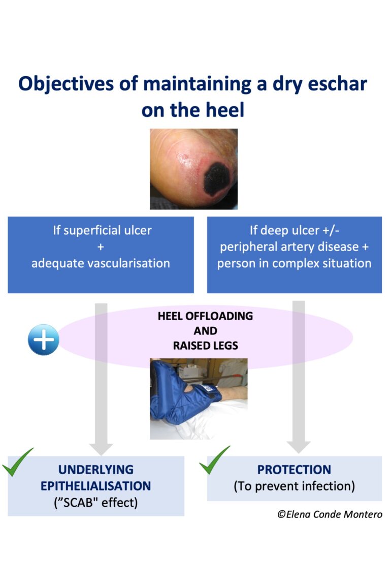
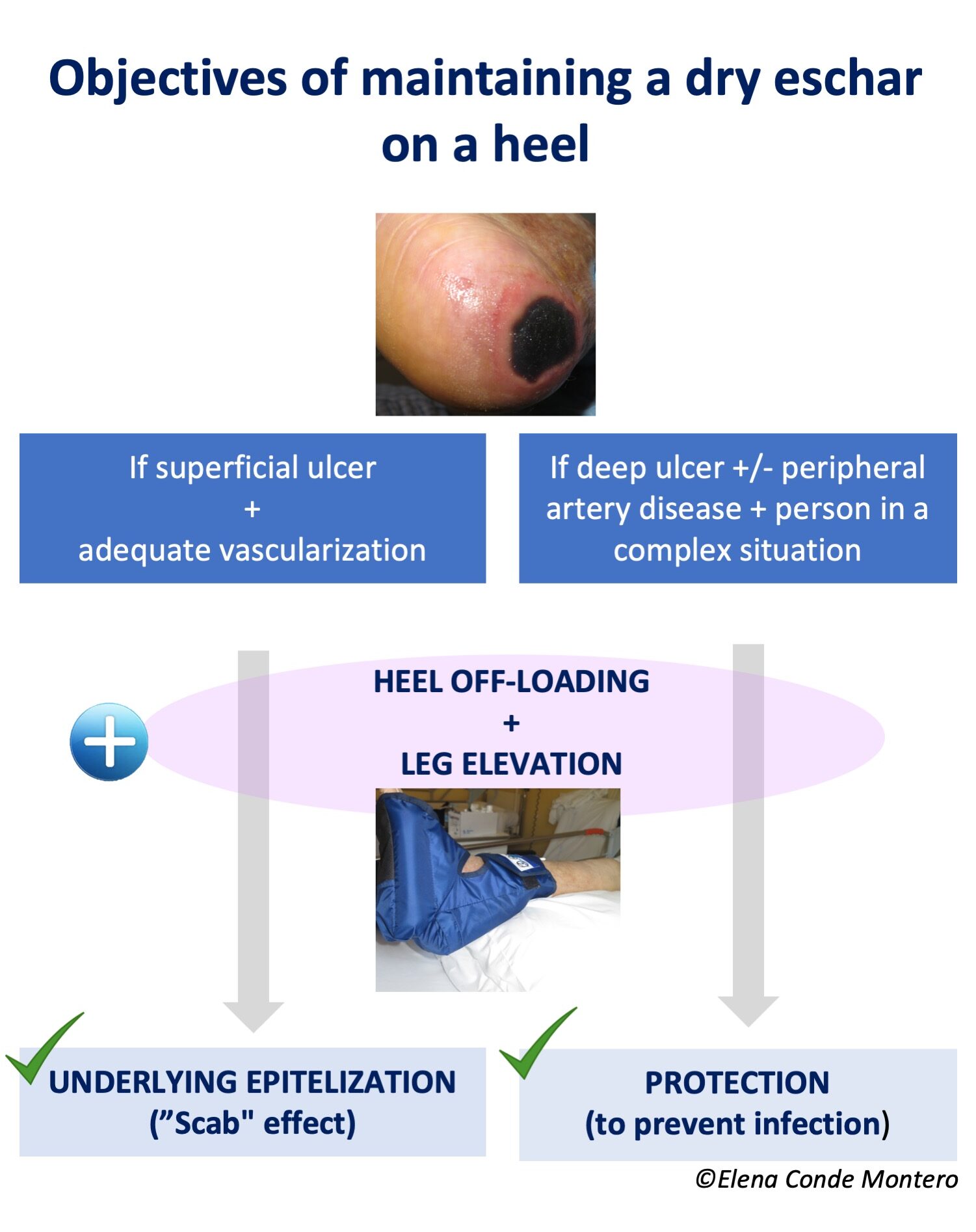
Because when they are dry, adherent to the wound bed, without fluctuation or other signs of infection, they have a protective function. Keeping this protective plaque dry can prevent bacterial growth and have a “scab effect”. It also avoids the pain of sharp debridement and the possibility of exposing deep tissues (calcaneus) with potential for superinfection.

If the ulcer is superficial, not extensive and vascularisation is adequate, this scab could promote underlying epithelialisation. On the contrary, if the ulcer is deep and extensive, there is associated peripheral arterial disease and/or the complexity of the patient’s situation does not allow another approach that favours healing, the objective will be exclusively to maintain the “protective scab” to avoid superinfection (palliative protection or prior to revascularisation or amputation).

How do I keep these necrotic plaques dry?

With astringent and antiseptic products and avoiding anything that provides moisture.

If the scab is completely dry and adherent, the strategy is to leave it open air.

To promote drying, povidone iodine (see post: “Povidone iodine and chronic ulcers: a great controversy”) or calcium alginate fibre dressings for their “scab” effect (see post: “Why do we use so many alginate fibre sheets in our wound practice?”) are good alternatives.

DON’T FORGET THAT THE MOST IMPORTANT THING IS TO PREVENT CELL DEATH FROM PROGRESSING, SO PRESSURE RELIEF WITH HEEL PRESSURE RELIEF AND LEG ELEVATION WITH HEEL SUSPENSION ARE ESSENTIAL. IN ADDITION, IF THE MAIN CAUSE IS ARTERIOPATHY, THE VASCULAR SURGEON WILL ASSESS THE POSSIBILITY OF REVASCULARISATION.

Happy summer!

Keratinocytes and their analgesic effect

I have decided to dedicate this post to the specific analgesic potential of keratinocytes because it is something that is hardly talked about in the world of wound healing and yet it is observed daily in clinical practice by all of us who graft wounds:)

In several blog posts I talk about the pain reduction we achieve when grafting painful wounds. In fact, one of them is directly entitled “Punch grafting as a painkiller for painful ulcers”  in which I discuss the results of a prospective study in which we included 136 patients with wounds of different aetiologies, in whom we measured the intensity of pain, with a visual analogue scale (VAS), before punch grafting (t0) and in the following 3 weeks visits (t1,t2,t3).  The primary endpoint was pain reduction. We found that pain reduction was clinically and statistically significant throughout the time of registration and, strikingly, we found no correlation between the percentage of graft attachment and pain reduction.

A sentence I repeat in these posts (also in classes or lectures I give) is: “Even if not all punch grafts do not adhere to the wound bed, they release growth factors and cells that promote healing and reduce pain”.

And why don’t I give more details about the exact mechanism of pain reduction? Because they are not known… In fact, I have not found any articles that analyse it… However, I have found evidence on the general role that keratinocytes may play in pain modulation. Although studies are not specific to grafts and in the reduction of pain with punch grafts there are many more factors potentially involved (fibroblasts, protein matrix, other proteins and growth factors), I think it is important to talk about the specific role of the keratinocyte, since using epidermal grafts (See post: “Differences between epidermal and dermo-epidermal punch grafts”) or keratinocyte cultures there is also evidence of pain reduction.

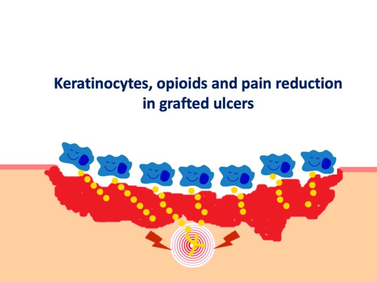
Available evidence suggests that keratinocytes can communicate with the nervous system via the opioid receptor system. Indeed, studies have shown that cells such as keratinocytes and fibroblasts play an important role in endogenous peripheral antinociception, as they are able to synthesise and secrete peptides such as enkephalins, a type of opioid neurotransmitter. The release of proinflammatory cytokines, especially IL-1b, induces the expression and release of opioids by these cells, which, once bound to receptors on peripheral nerve fibres, inhibit the release of pain neurotransmitters such as substance P.1,2.

A recent study has shown that keratinocyte and fibroblast culture supernatant promotes analgesia during inflammatory pain induced in a rat model of hyperalgesia, corroborating previous studies. In this study, administration of a non-selective opioid antagonist (naloxone) inhibited the antihyperalgesic effect of keratinocyte culture supernatant, demonstrating that the effect of keratinocyte culture supernatant is mediated by the release of opioids.3

Thus, the release of endogenous opioids by keratinocytes, stimulated by the pro-inflammatory wound environment, may be one of the pathways by which punch grafts have such striking analgesic power.

An exciting topic for further research!


Emollients for eczema on legs

What is the best moisturising recommendation for the elderly with dry skin or those with eczema secondary  to venous insufficiency?

You have probably asked yourself this question many times. However, while dry skin and atopic dermatitis in children are much talked about (and many products have been marketed for their treatment), research on the use of emollients in older people and stasis eczema is very scarce.

I was encouraged to write this post after reading this recent Cochrane review: “Hygiene and emollient interventions for maintaining skin integrity in older people in hospital and residential care settings”  Although skin dryness and fragility, as well as pruritus, are very prevalent in older people (and have a huge impact on quality of life), the main conclusion of this meta-analysis is that more studies are needed to make recommendations on the use of emollients and hygiene products to protect and repair the skin barrier in older people.

As I discussed in the post “Eczema craquelé: when skin deteriorates like paintings”, in older people, the stratum corneum (the most superficial layer of the epidermis) does not have an adequate barrier function, with a consequent increase in permeability that will lead to inflammation and itching. Dry skin lesions occur particularly on the legs. So, although little discussed, considering eczema due to dryness and eczema due to venous hypertension, leg eczema in adults and older people competes in frequency with atopic dermatitis in children.

Although more studies are needed in older people and people with venous insufficiency, the use of emollients seems essential on a preventive and therapeutic level whenever we encounter dry skin or eczema.

To understand this need for the use of emollients, we first need to understand the physiology of the skin barrier (I recommend the article Use of emollients in dry-skin conditions: consensus statement).

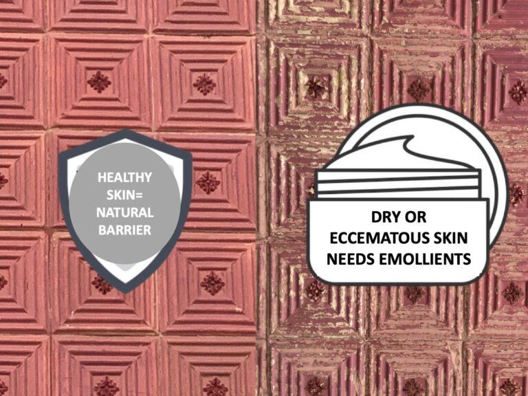
The stratum corneum (outermost layer of the epidermis) forms a barrier that restricts water loss and prevents the penetration of irritants and allergens. It is composed of well hydrated and compact corneocytes (degenerated keratinocytes) that are surrounded by a matrix of intercellular lipids (ceramides, cholesterol and fatty acids). The corneocytes contain the natural moisturising factor, a set of hygroscopic compounds that attract and retain water in the cells. Together, these natural moisturisers are necessary to maintain skin plasticity and prevent dehydration of the corneocytes, which would lead to cracking. Both the lipid-filled extracellular compartment and the humectant-filled intracellular compartment are essential for the barrier function and water retention of the stratum corneum, as well as maintaining a low pH. In other words, it is “a wall of bricks held together by cement”. Soaps and detergents remove lipids, strip the natural moisturising factor and increase the pH, so it is essential to use syndet cleansing products.

In contrast to creams that only provide hydration, emollients also prevent water loss. Therefore, we will always recommend emollients in case of diseased skin (dryness, eczema, psoriasis, other dermatitis).

Simple emollients leave a thin occlusive layer of non-physiological lipids or oils, such as petroleum jelly, on the surface of the skin, thus reducing water loss from the stratum corneum. Emollient products can be formulated with additional ingredients such as humectants, physiological lipids and antipruritic agents. Humectants, such as urea and glycerol, attract and retain water in the stratum corneum, thereby compensating for reduced levels of natural moisturising factor and other natural humectants in diseased and ageing skin. Physiological lipids, such as ceramides, cholesterol and free fatty acids, replenish and restore the intercellular lipid matrix.

Not all emollients are the same, so the choice of the best product will depend on the needs of each individual’s skin. In addition, for effective protection of the stratum corneum, we should also use cleansing products with emollient action, short showers and avoid aggressive rubbing drying (see more recommendations in the post: “Eczema craquelé: when the skin deteriorates like paintings”).

Proper use of emollients can have a direct impact on the quality of life of all people with dryness or eczema on legs, by reducing disabling symptoms such as itching and reducing outbreaks of dermatitis and the need for topical corticosteroids.

Eczema craquelé: when skin deteriorates like paintings

The craquelé effect on the skin is often found on the legs of the elderly, especially in winter. The name of these lesions, eczema craquelé, is very intuitive, but not very well known among professionals who treat leg ulcers. So, let’s dedicate a post to understand why it occurs, identify how it presents itself and how to prevent and treat it.

What is eczema craquelé?

Eczema craquelé, also known as asteatotic eczema, is characterised by the presence of superficial, criss-crossed fissures on a more or less erythematous and scaly base, with the subsequent appearance of craquelure or cracked pavement. It is usually very itchy.

The extent of the lesions is variable and scratching may aggravate the inflammation and lead to erosions or ulcers.

As mentioned above, it usually affects the lower limbs of older people.


Why does it occur?

It has been classically associated with xerosis (dry skin) and factors that alter the barrier function of the stratum corneum (the most superficial layer of the epidermis), such as age, the use of soaps, long showers with hot water or a cold, dry environment.

Old people have reduced activity of the sebaceous and sweat glands. In addition, age-related alteration of the defensive functions of the stratum corneum leads to increased permeability which will cause inflammation and pruritus.

However, this crackling is also observed in cases of rapid onset oedema, such as decompensation of heart failure, hypoproteinaemia or phlebolymphoedema. In fact, the distension of the skin secondary to these causes of rapid oedema is the cause of the production of “fractures” in the stratum corneum and the rest of the epidermis. It is therefore possible that even in the classic presentation of eczema craquelé (old person with xerosis) the preference for the legs may reflect the frequent occurrence of oedema due to venous hypertension in this age group. Moreover, the term “acute oedema-cutaneous distension syndrome” has been proposed to include this “craquelé” presentation in the spectrum of skin lesions associated with rapid onset oedema. Depending on the elasticity of the skin, the person will respond to this oedema with blisters or with eczema craquelé. 2

This photo shows a case of eczema craquelé in the context of a flare-up of phlebolymphoedema.

On the other hand, in addition to the typical presentation, eczema craquelé can also occur in other locations, such as on the trunk or generalised over the whole body, and in younger people. In these cases, as it may be the expression of an underlying systemic disease, such as a malignancy, a detailed study must be carried out.3

How can it be treated and prevented?

Prevention is just as important as treatment. Bearing in mind that xerosis is the main trigger for these lesions on legs, the strategy will always be focused on promoting skin hydration and avoiding external factors that damage the skin’s hydrolipidic film. Therefore, these are the main recommendations, which should be explained to the patient and family members:

  • Lower the water temperature and shorten the shower time.
  • Avoid the use of soaps
  • Apply emollients several times a day, especially after bathing.
  • Use humidifiers
  • Use cotton clothing and avoid wool.
  • Avoid scratching
  • Control of oedema if it is the trigger (therapeutic compression, anti-oedema measures).
  • In case of extensive, very itchy lesions, which are not controlled with the previous measures, use topical corticosteroids of medium-high potency. Ideally, it should be mixed with moisturiser and applied to damp skin after showering and immediately put on cotton pyjamas. Use it for 1 to 2 weeks, depending on the evolution. Calcineurin inhibitors are an alternative to topical corticosteroids.4
  • Oral antihistamines can be added to control pruritus.

References:

  1. Yang CS, Lott JP, Bunick CG, Bolognia JL. Eczema craquelé associated with nephrotic syndrome. JAAD Case Rep. 2016;2(3):241-243.
  2. Cox, Neil, Chalmers, Robert, MB, FRCP, Bhushan, Monica. The Acute Edema/Cutaneous Distension Syndrome. Arch Dermatol. 2003;139(2):224-225. 
  3. Sparsa A, Boulinguez S, Liozon E, Roux C, Peyrot I, Doffoel-Hantz V, Labrousse F, Vidal E, Bordessoule D, Bonnetblanc JM, Bédane C. Predictive clinical features of eczema craquelé associated with internal malignancy. Dermatology. 2007;215(1):28-35.
  4. Schulz P, Bunselmeyer B, Bräutigam M, Luger TA. Pimecrolimus cream 1% is effective in asteatotic eczema: results of a randomized, double-blind, vehicle-controlled study in 40 patients. J Eur Acad Dermatol Venereol. 2007 Jan;21(1):90-4.

The importance of footfall and footwear in venous ulcers

How little is said about the importance of footfall and footwear in people with venous ulcers! In this post we will understand why it is important to raise awareness of their relevance.

As we saw in the post, “Chronic venous insufficiency from a dermatological perspective” , the failure of the muscular pump is one of the triggers of venous hypertension, which is the cause of all the skin alterations framed in chronic venous insufficiency.

In addition to calf muscle activation, the plantar pump is essential to reduce ambulatory venous hypertension and thus promote venous ulcer healing.

There is increasing awareness of the need for optimal therapeutic compression, to increase the effectiveness of calf muscle contraction, but we do not give enough importance to footwear and effective stride to activate the plantar pump.


Although there are few studies on the subject, those that have been published suggest that alterations in foot statics may be an important, but underestimated, risk factor for chronic venous disease.

Anatomical studies of the foot pump indicate that the venous reservoir is located in the lateral plantar veins. When the sole of the foot presses against the ground, the lateral plantar veins empty and blood return is promoted by the posterior tibial veins and the internal saphenous system via the medial perforators of the foot. Therefore, the presence of alterations in foot statics could be a good explanation for impaired venous return during ambulation.1 In addition, inadequate footing leads to postural changes that may reduce the efficacy of the calf muscle pump.

In a retrospective study of 412 patients (824 feet), seen by the same phlebologist, disorders in foot statics were recorded. 62.2% were women and 59.3% had a CEAP classification of C3 or higher. Thirty-one per cent had some foot statics disorder (16.6% pes cavus and 14.5% flat feet), but from C2 onwards the prevalence rose to 38%. A statistically significant association was found between the severity of venous disease and the presence of these alterations .2

Another prospective study of 24 patients with symptomatic chronic venous disease and impaired foot statics (mostly women with pes cavus), shows how correction of foot motion impairment with an insole can have comparable effects to a therapeutic compression stocking (18 mmHg) on quality of life and symptoms. In fact, the authors suggest that many symptoms of patients with varicose veins (pain, heaviness, cramps) probably do not have a venous origin, but rather postural alterations secondary to abnormalities in the footprint. They therefore recommend a biomechanical assessment of the foot in all patients with chronic venous disease and, if necessary, the use of insoles associated with compression therapy to optimise the efficiency of the veno-muscular pumps from the foot to the hip during gait and, therefore, improve postural problems and combat venous hypertension.3

On the other hand, if it is so clear that venous return begins at the sole of the foot and the proper functioning of this pump is key, why do patients continue to leave the clinic with a shoe that does not fit the bandage, that displaces it or that does not allow them to walk normally?

Taking all this into account, especially the frequency of alterations in the gait of these patients and their potential impact, the practical recommendations for people with venous ulcers should be as follows:

  • As long as bandages are used, shoes should be wide, with a fastening that adapts to the foot, providing comfort and stability during walking.
  • To prevent recurrence, in addition to a compression stocking, it will be important to use a suitable shoe and a biomechanical study of the foot carried out by a podiatrist in case an insole is necessary.

A subject that needs, without a doubt, more studies …I would love podiatrists to share their opinion and experience on this subject!:)

References:

  1. Uhl J-F, Gillot C. The foot venous pump. Anatomy and physiology. Phlebolymphology 2010;17:151– 8
  2. Uhl JF, Chahim M, Allaert FA. Static foot disorders: a major risk factor for chronic venous disease? Phlebology. 2012 Feb;27(1):13-8.
  3. Uhl JF, Chahim M, Allaert FA. Compression versus inner sole for venous patients with foot static disorders: a prospective trial comparing symptoms and quality of life. Phlebology. 2015 Feb;30(1):32-8. 

Winter’s famous article and moist wound healing

We are going to analyse probably the most referenced article about wound healing, but probably also the least read.

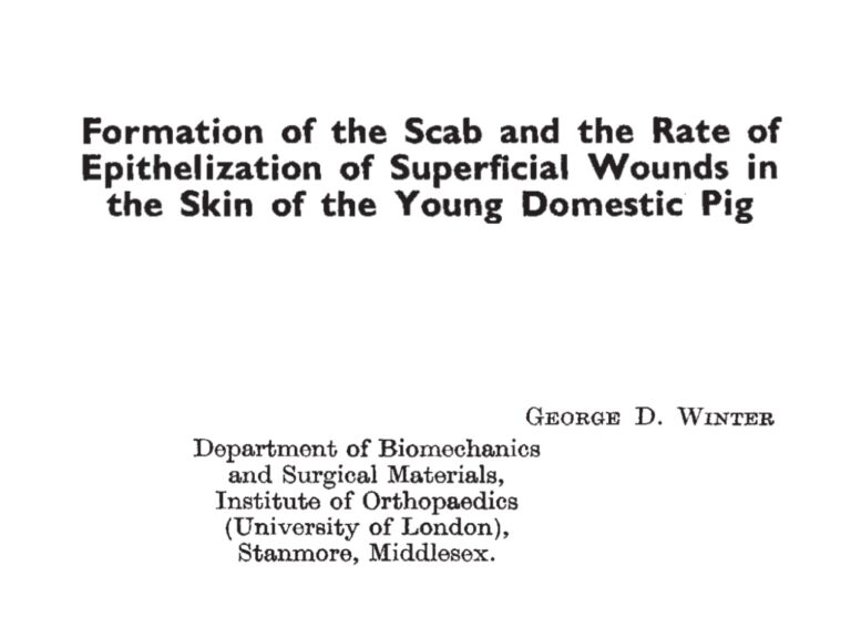
In fact, in any scientific text that talks about “moist wound healing” in chronic ulcers, you find the reference “Winter GD. Formation of the scab and the rate of epithelization of superficial wounds in the skin of the young domestic pig. Nature. 1962 Jan 20;193:293-4″.

It seems to be universally accepted that this article by George Winter marks a “before and after” in the world of wound healing… If until then it was thought that the best thing to do was to dry the wounds, following this study “moist wound healing” began to be talked about as the cornerstone of modern wound treatment and, in parallel, the boom in dressings.

If you haven’t read this very famous (and very short) article, you might be surprised by these details of its methodology and results, as well as Winter’s actual conclusions. Here we go:

  • It’s an experimental study with pigs, young and healthy. It is not clear how many pigs were included, but it only mentions two.
  • Wounds were created on the back up to the papillary dermis, i.e. clean superficial surgical wounds.
  • The area where the wounds were made had been waxed beforehand.
  • To compare the estimated epithelialisation rate between wounds covered with a polyurethane film (“moist wounds”) and uncovered (open air) wounds, serial skin biopsies were performed.
  • Each pig was used as its own control. Twelve wounds were studied (6 in each group).
  • In the results the author highlights that, at day 3, the amount of new epidermis (observed under the microscope) was twice as high in the “moist” wounds as in the uncovered ones, i.e. a higher rate of epithelialisation. However, although it does not say so in the text, the table in the article shows that from day 7 onwards there is no difference in the rate of epithelialisation.
  • He also suggests a faster formation of connective tissue in “moist” wounds, but the article does not provide any data to support this.
  • Winter concludes that “the demonstration that a simple change in physical conditions at the wound surface can have such a marked effect on the rate of epithelialisation is highly relevant to experimental methods in wound healing. It would be imprudent to draw conclusions about the specific effects of different products on the rate of healing”.

Were you surprised? Well, this article with 12 acute superficial wounds in pigs is the starting point of the spiral of generalisation of the “moist environment” in chronic wounds…

I would like to point out that in the discussion of his article Winter mentions the role of the scab in preventing the entry of dirt and microorganisms, as well as protecting the newly formed epidermis from dehydration. In our practice we perform daily wounds with the same characteristics as in Winter’s study, down to the papillary dermis, to obtain punch grafts… And 60 years after this article no dressing has been shown to be superior to the rest, or even to scabs, in clean superficial acute wounds! See entries: “Which dressing should I choose to cover the skin graft donor site?”  and “Tricks to avoid complications in acute wounds”.

As you already know, what I like to talk about is “optimal environment”… Which is still not clear what it is… So I am going to finish by mentioning an article that, contrary to Winter’s, is very little referenced… The author makes a very interesting reflection, which I cannot share more:

“Since we have been using scabs as wound coverings for thousands of years, we should try to improve their performance, instead of ignoring the benefits of this very effective dressing” Nelson EA. Moist wound healing: critique II. J Wound Care. 1995 Sep 2;4(8):370-371.

In fact, when I read this reflection, my first thought was: “Wow… And I thought this sentence was mine ;)”.

Repositioning and not removing scabs: two unwritten rules of punch grafting

The truth is that there are few written rules about punch grafting… In fact, it is difficult to find scientific publications with the most relevant practical tricks to succeed with this technique… To make up for this deficit, punch grafting is the star topic of this blog, but I have never talked in detail about these two essential points. Both “repositioning the grafts” and “not removing the scabs” are in line with the recommendations “do not touch” and “maintain the pro-healing microenvironment” that I always talk about. Let’s understand the reason for these two rules:)

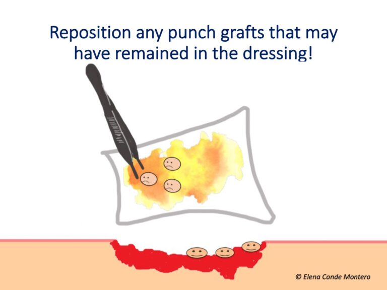
Replace the grafts that have remained in the dressing!

It is not uncommon to find a punch graft in the dressing when the dressing is removed.

Several situations can occur:

  • Absence of attachment: grafts in which no new vessels have formed to nourish them from the wound bed remain yellowish and swollen due to imbibition (due to the nutrition that has reached them through contact with the wound exudate). This usually occurs if the wound bed is not optimal or if there is not adequate local fixation and rest during the first 3-4 days.
  • Pulling out: the grafts have taken hold, they have a pinkish-bluish colour, but we have “pulled them out” when removing the dressing.
  • Detached epidermis: the grafts are adhered to the bed, pink, shiny, but their most superficial layer (transparent sheets of keratinocytes) have stuck to the dressing.

What is the best dressing to prevent the grafts from detaching? No comparative studies have been done, but our experience is good with the use of interphases or alginate fibre sheets directly on the wound (which, if it is very adhered, should be moistened before removal).

How do we recover and reposition them? We will use tweezers to separate them from the dressing and we will use the tip of a scalpel or needle to spread them out again on the wound bed.

What if we are not sure which side corresponds to the dermis and the epidermis? A clue to differentiate between them is that the dermis is shinier. However, even if we do not know which side is which, and we place them the other way round, this will also favour healing.

Should we recover the grafts even if there is a lot of exudate in the dressing? Yes, always, although sometimes it is not easy to find them 😉

Do not remove scabs!

As in acute wounds with a good evolution, in grafted wounds, scabs will form in parallel to epithelialisation. These scabs, consisting predominantly of dehydrated layers of cellular debris and exudate, act as a protective barrier to maintain a healing-promoting microenvironment. In fact, to prevent excess moisture in the grafted wound and encourage these scabs to form, we usually apply zinc oxide solution in combination with alginate fibre sheets (see post “Why do we use topical zinc on wounds and perilesional skin?” and “Why do we use so many alginate fibre sheets in our wound practice?”

Scabs should only be removed if they fluctuate, if purulent exudate comes out on pressure or if there are other signs of infection. They will fall off when they are no longer necessary… At that time, we can help them to fall off by moisturising them, for example with pure petroleum jelly 😉


How long can I space out dressing changes of a leg ulcer?

First the pandemic and now the big snowfall… This year we have accumulated impediments to maintain the healing guidelines that we consider most beneficial for our patients… Here are some questions that you may have asked yourself at some point in the current times:

How long can I keep this dressing and dressing untouched?

What happens if it goes on for more than a week?

Is there a time limit that must not be exceeded?

This post will solve all your doubts about the frequency of wound dressings;)

The most important idea, and one that we must be very clear about, is that the frequency of dressings will depend on the evolution1…. If the wound has exudate that is difficult to control, abundant biofilm or signs of infection, dressing changes must be frequent to encourage the removal of bacteria, proteases and other factors that hinder healing. Dressing should also be more frequent if we want to closely monitor the wound’s response to a treatment that requires supervision, such as the initiation of compression therapy in special situations.

However, if the evolution is favourable, dressing changes should be spaced as far apart as possible. The limit of this spacing will be set by the control of exudate, pain, itching and the maintenance of the dressing in place. In other words, there is no set limit and the times are determined by the evolution of each patient.


So, can we space out dressing changes by 15 days? If these factors are met, we can do it and we must do it. We must not forget that in each dressing change we remove denatured tissue and exudate that is negatively affecting healing, but we also remove cells, growth factors and other proteins that promote wound closure. In addition, at dressing changes, the temperature of the wound bed decreases, with the consequent inhibition of mitotic activity, which takes time to restart (See post: “The art of wound cleansing”).

And more than 15 days? Well, as we have said, it could be, it depends on the case. In fact, sometimes the dressing is kept indefinitely to promote epithelialisation with a “scab effect” and only the edges of the dressing are removed and gradually peeled off (for example, alginate dressings in superficial acute wounds). See post: “Why do we use so many alginate fibre sheets in our wound practice?”

Therefore, the ideal is to select a primary and secondary dressing with characteristics that allow adequate exudate control and, in addition, a bandage that lasts over time with adequate pressure and adapts to the needs of each patient. If the only problem is that the bandage falls off or is displaced every few days (e.g. due to oedema reduction or patient mobility), we can only change the bandage and space the dressing changes. In these cases, in small wounds,  wrap compression devices with velcro fasteners are a very interesting alternative, as they allow daily self-adjustment, maintaining the necessary pressure. See post: “Why are adjustable compression wrap devices so popular?”

In short, let’s be flexible in the frequency of dressing changes and, if the wound is going well, let’s remember that “less is more”.