Home Blog Page 9

Have you ever heard of the topical use of sevoflurane in wounds?

If you have never heard of this alternative treatment for painful ulcers, surely while reading this post you may think of more than one patient who could benefit from its use.

Once again, Carmen Alba Moratilla’s scientific curiosity and motivation have been important driving forces in my contact with this therapeutic alternative, which I have been able to put into practice thanks to the collaboration of Dr. Afredo Abad, head of the Anaesthesiology Service at the Infanta Leonor University Hospital. Our experience is recent and therefore very limited, but the findings are in line with those described by other authors, which we will now sees.

Pain is one of the main factors limiting the quality of life of people with chronic leg wounds and, in many cases, a therapeutic challenge for healthcare professionals. Fear of pain during treatment can trigger anticipatory anxiety, which causes much suffering in these patients. Before trying different analgesic drugs, the first thing to do is to identify the cause of the pain, especially if it has increased sharply, as it may be a sign of infection, inadequate compression, with excessive pressure points or poor control of oedema and exudate, or contact dermatitis due to the dressings used. Although any leg ulcer, regardless of its cause, can be painful, the type of pain can guide us in the etiological diagnosis.

The side effects inherent in systemic analgesics complicate decision-making on the best treatment strategy, especially in older patients or those with multiple comorbidities. Although dressings with a potential topical analgesic effect are being developed and marketed, in our clinical practice the range of products available is very limited and is usually reduced to lidocaine/prilocaine cream (EMLA), whose analgesic efficacy during sharp debridement has been proven in different clinical trials.1 Pharmaceutical compounding allows us to transport morphine in a hydrogel for topical application. However, although the analgesic effect of topical morphine in wounds has been described in case series, the few clinical trials performed, with small sample size among other limitations, do not detect statistically significant differences in comparison with placebo.2 On the other hand, the analgesic effect of advanced therapies such as platelet-rich plasma or punch grafting has been described. These therapies are especially interesting in Martorell hypertensive ischemic ulcer, which is enormously painful.

In fact, my first contact with topical sevoflurane has been in a patient with Martorell ulcers in both legs, with tendon and bone exposure, with uncontrollable despite treatment with morphine and gabapentin. This first experience could not have been more successful, since after weekly applications for a month, with excellent analgesic control from the first session, we were able to apply punch grafts on the wound and achieve complete epithelialization. Well, let’s go to what interests you: What is sevoflurane and how does it work in wounds?

Sevoflurane is an inhalation anaesthetic agent, of the group of halogenated ethers,  for use in induction and maintenance of general anesthesia. It is a colourless volatile liquid, packaged in 250 ml bottles, which is vaporised for use as an anaesthetic gas. Although it has traditionally been assumed that the analgesic action of halogenated anaesthetics is exclusively central, they are also known to have an analgesic action at the peripheral level. This concentration-dependent effect is due to the fact that, when applied locally, sevoflurane can reach sufficient partial pressure to block peripheral nociceptors. It can therefore be used off-label in its liquid form, as a local anaesthetic, applied by irrigation to painful wounds.3

Although studies published in the literature are mostly isolated cases or short series, the results are promising.3-6 The benefit of topical sevoflurane seems not to be limited to its analgesic effect, which appears almost immediately and makes effective sharp debridement possible. It has been proposed that it can produce a healing acceleration due to its vasodilator effect and, although its mechanism of action is unknown, bactericidal action has been detected in vitro against multidrug resistant strains of S. Aureus, P. Aeruginosa and E.Coli.7

The benefit/risk profile is very favourable since, as side effect, only itching and perilesional erythema have been described after application. Although sevoflurane blood levels have not been measured, no systemic effects have been recorded after local use.

The first published case series6 includes 9 patients with painful venous ulcers resistant to systemic analgesic treatment, in which topical sevoflurane was used. The average number of dressing changes per patient was 8 (range: 4-13), but the authors do not specify the frequency of dressing changes. Irrigation of the wound bed was performed with approximately 1 ml/cm2 of liquid sevoflurane, always after cleansing with physiological serum. All patients experienced a rapid reduction in pain at rest (less than 2 minutes), which was maintained over time, an average of 10 hours. This reduction in pain, measured by a verbal pain scale (0-10) was clinically and statistically significant in all applications (average pain reduction of 6 points). Complete wound healing was obtained in 4 patients.

Other Spanish authors5 confirm the benefit of topical sevoflurane application in painful recalcitrant wounds. They reported 36 patients with excellent pain control in 94% of cases, immediately after sevoflurane application and maintained over time (variable duration, between 2 and 48 hours). They found no loss of efficacy over time (length of treatment ranged between 3 and 24 months).

The results of a randomised clinical trial8 designed to assess the impact of topical sevoflurane use on pain, daily opioid dose and wound size have recently been published. In the control group (conventional management), 10 patients with leg ulcers were included, while 5 were assigned to the treatment group. The frequency of sevoflurane irrigations (1ml/cm2) ranged from 1 to 4 per day, with the aim of maintaining a pain score (VAS pain) inferior to 3. Treatment was performed for 90 days. Among the limitations of the study it must be highlighted the small sample size, the different basal characteristics between groups and the variability inherent in the application of the product by different people (health professionals or the patients themselves). A statistically significant reduction in pain, opioids uptake and wound area was found in the treatment group. As a mild side effect, already described by other authors and which we have also observed, perilesional redness appeared in 4 of the 5 patients being treated with sevoflurane.

There are still many outstanding questions that will find answers over time, thanks to experience and the conduct of more experimental studies. These include the following: what dose and frequency of application would be optimal, what type of wound would benefit most from treatment, what safety measures should be required for its use?

Do you have any experience with topical sevoflurane?


References:
1. Briggs M, Nelson EA, Martyn-St James M. Topical agents or dressings for pain in venous leg ulcers. Cochrane Database Syst Rev. 2012 Nov 14;11:CD001177. 
2. Bastami S, Frödin T, Ahlner J, Uppugunduri S. Topical morphine gel in the treatment of painful leg ulcers, a double-blind, placebo-controlled clinical trial: a pilot study. Int Wound J. 2012;9(4):419-27. 
3. Lafuente-Urrez RF, Gilaberte Y. Sevoflurane: a valid alternative for the treatment of vascular ulcers? Actas Dermosifiliogr 2014;105:202–3. 
4. Imbernón A, Blázquez C, Puebla A, Churruca M, Lobato A, Martínez M, Aguilar A, Gallego MA. Chronic venous ulcer treatment with topical sevoflurane. Int Wound J.2016;13(5):1060-2. 
5. Dámaso Fernández-Ginés F, Cortiñas-Sáenz M, Navajas-Gómez de Aranda A, Yoldi Bocanegra R, Sierra-García F. Reply: to Chronic venous ulcer treatment with topical sevoflurane by Imbernón et al. Int Wound J. 2017;14(3):591 
6. Gerónimo Pardo M, Martinez Serrano M, Martínez Molsalve A, Rueda Martínez JL. Usos alternativos del sevoflurano. Efecto analgésico tópico. Rev Electron Anestesia 2012; 4:181.
7. Martínez M, Gerónimo M, Crespo MD. Actividad bactericida del sevoflurano frente a Staphylococcus aureus, Pseudomonas aeruginosa y Escherichia coli. Enferm Infecc Microbiol Clin 2009;27:120–1.

8. Dámaso Fernández-Ginés F, Cortiñas-Sáenz M, Mateo-Carrasco H, de Aranda AN, Navarro-Muñoz E, Rodríguez-Carmona R, Fernández-Sánchez C, Sierra-García F, Morales-Molina JA. Efficacy and safety of topical sevoflurane in the
treatment of chronic skin ulcers. Am J Health Syst Pharm. 2017;74(9):e176-e182. 

Punch grafting and negative pressure therapy: a successful couple

In previous posts we have talked about the benefits that these two treatments have, separately, promoting healing. However, their combined use plays a fundamental role. On the one hand, their sequential use is of great interest, as the application of negative pressure promotes the formation of granulation tissue, with reduction of exudate and bacterial load, and therefore prepares the wound bed to facilitate graft taking. On the other hand, its synchronous use can improve the percentage of graft taking,1-6 especially in certain situations that we will now point out.

As I commented in previous posts, among them “Types of skin grafts to cover chronic wounds: which one to choose“, in our wound clinic we normally use punch grafts. These partial thickness grafts are dermo-epidermal fragments more or less circular or oval, which are usually obtained with punch (punch), scalpel or curette, without deepening beyond the papillary dermis. The procedure is performed under local anaesthesia in the donor site, usually the thigh. The fragments are placed directly on the wound bed, without any type of fixation, apart from pressure and local immobilization during the first days after the procedure.

Most authors of the published studies about the benefits of negative pressure therapy in skin graft taking, use mesh skin grafting, which is another type of partial thickness graft.3-5 However, a clinical trial involving 60 patients with chronic leg ulcers treated with punch grafts finds a statistically significant reduction in healing time, pain and treatment costs in the group that received associated negative pressure therapy.6

The major determinant of the success of skin grafting is its attachment to the wound bed. It also represents the greatest challenge, especially in chronic wounds in particular anatomical locations (Achilles tendon, ankle), where graft attachment to the wound bed is difficult. On the other hand, we cannot forget that advanced age is a constant feature in patients with wounds and, therefore, so is skin fragility (dermatoporosis). The pressure that must be exerted on the grafts to favour graft taking (and the friction of the material used) can produce the opposite effect in patients with skin ageing, as it can damage capillaries and trigger bleeding and haematomas. In addition, although the ideal situation is to graft wounds with optimal granulation tissue, our experience tells us that this is not easy to achieve with chronic ulcers of long evolution in elderly patients with multiple comorbidities. Conclusion: the majority of wounds that we graft do not present a perfect microenvironment for the reception of those skin fragments. Although grafts that do not take are also interesting because they release growth factors and other molecules and cells that promote healing, the more percentage of graft taking achieved, the better.

There are different negative pressure therapy devices commercially available on the market. We use a portable, single-use device with a pump that exerts a pressure of -80 mmHg on the dressing that we place on the wound covered with the grafts.

A. Recalcitrant post-traumatic ulcer, 4 month-long, in Achilles tendon. B. Coverage with seal grafts. C. Clinical aspect in the first dressing change, at week one after the procedure (punch grafting + negative pressure therapy) D. Complete epithelialization at week 7.

Our experience, like that of other professionals, confirms the benefit of applying negative pressure therapy on skin grafts. Its efficacy is associated, on the one hand, with a reduction in exudate and better immobilization and sealing of the grafts, with the consequent decrease in shear and, therefore, lower risk of development of seromas and hematomas.1-6 On the other hand, it has been proposed that the mechanical stretching that produces negative pressure can both stimulate the signaling pathways that promote keratinocyte mitosis and activate neo-angiogenesis by increasing microcirculatory flow in the wound bed and edges.2

I cannot finish a post that includes the word “successful” in the title without referring to the underlying engine of every successful idea and treatment strategy in our wound clinic: I AM PART OF A WONDERFUL TEAM.


Vicente Soldevilla


References:

  1. Gupta S. Optimal use of negative pressure wound therapy for skin grafts. Int Wound J. 2012;9(Suppl 1):40–7. 
  2. Azzopardi EA, Boyce DE, Dickson WA, Azzopardi E, Laing JH, Whitaker IS, Shokrollahi K. Application of topical negative pressure (vacuum-assisted closure) to split-thickness skin grafts: a structured evidence-based review. Ann Plast Surg. 2013 Jan;70(1):23-9.
  3. Moisidis E, Heath T, Boorer C, Ho K, Deva AK. A prospective, blinded, randomized, controlled clinical trial of topical negative pressure use in skin grafting. Plast Reconst Surg 2004;114:917–22. 
  4. Llanos S, Danilla S, Barraza C, Armijo E, Pineros JL, Quintas M, et al. Effectiveness of negative pressure closure in the integration of split thickness skin grafts. A randomized, double-masked, controlled trial. Annals of Surgery 2006;244(5):700-5. 
  5. Maruccia M, Onesti MG, Sorvillo V, et al. An Alternative Treatment Strategy for Complicated Chronic Wounds: Negative Pressure Therapy over Mesh Skin Graft. BioMed Research International. 2017;2017:8395219. 
  6. Vuerstaek JD, Vainas T, Wuite J, Nelemans P, Neumann MH, Veraart JC. State-of-the-art treatment of chronic leg ulcers: A randomized controlled trial comparing vacuum-assisted closure (V.A.C.) with modern wound dressings. J Vasc Surg. 2006 Nov;44(5):1029-37 


Brief overview of wound healing

Healing is the natural process to regenerate the tissues that have suffered an injury. In the case of an erosion, which exclusively affects the epidermis, the tissue is restored, achieving a structure similar to the previous one. If, on the other hand, deeper structures are affected, this regeneration does not take place, and a scar occurs.

In acute wounds, there is a different healing mechanism depending on whether the lesions are of partial or total thickness. In partial thickness wounds, the annexes are preserved. These structures serve as a reservoir of epithelial cells and re-epithelialisation occurs from the edges and centre of the lesion. In full thickness wounds, since the annexes are not present, re-epithelialisation is only possible from the edges.

When a wound is produced, a series of complex biochemical phenomena occur in order to repair the damage. These phenomena occur with some temporal overlap but, for better understanding, they are consensually into the following phases: inflammatory, proliferative and remodelling phase.


Inflammatory phase:

This first phase consists of a vascular and cellular response. Platelets are the first cells to intervene, with their haemostatic role and their releasing of growth factors that stimulate healing. After an early vasoconstriction to stop bleeding, there is vasodilation, extravasation of fluid into the extracellular space and a blockage of lymphatic drainage, with the consequent appearance of cardinal signs of inflammation (heat, flushing and tumour). This first phase usually lasts 24-48 hours, but in some patients it can last up to 2 weeks. If it lasts longer, it is considered chronic inflammation and hinders healing.

When an injury occurs, the vessels are damaged and collagen is exposed. This collagen and the thrombin generated in the haemostasis process stimulate the platelets, producing their activation, adhesion and aggregation. Platelets release different mediators such as serotonin, thromboxane A2, fibrinogen, fibronectin and thrombospondine, thus keeping the aggregation process active to stop potential bleeding. The platelet-derived fibrinogen is converted to fibrin, which acts as a matrix for monocytes and fibroblasts. The release of prostacyclin by endothelial cells inhibits platelet aggregation, thus limiting the spread of the coagulation process. In this inflammatory phase, the role of growth factors, such as PDGF (platelet-derived growth factor), with chemothactic, angiogenic and mitogenic properties, is essential.

In the early hours of the inflammatory phase, neutrophils and monocytes are the predominant cells at the site of injury. Subsequently, their number decreases and the number of macrophages increases.


Proliferative phase:

In this second phase the cellular response predominates and it is the stage in which a permeable barrier is created (reepithelization), an adequate blood supply is established (revascularization) and a reinforcement of the damaged tissue takes place (fibroplasia).

Epithelialization is defined as the formation of epithelium over an area devoid of it. It involves the migration of epithelial cells from the edges of the wound to the centre. This process begins hours after tissue damage. The keratinocytes at the wound edges undergo structural changes, break their cell and basement membrane bonds, and are free to migrate over the wound surface, thanks to the formation of intracellular actin microfilaments. In partial-thickness wounds, adnexal structures that persist in the bed contribute to re-epithelialization.

Epidermal cells secrete collagenases and plasminogen activator. Plasmin production promotes clot dissolution and allows keratinocyte migration. When re-epithelialization is complete, epidermal cells recover their intercellular and basement membrane junctions. The moist environment encourages the re-epithelialization of wounds. Different studies show the benefits of occlusive healing to limit inflammation, restore barrier function and increase skin moisture (Mustoe, 2011).

Angiogenesis:

The migration and proliferation of endothelial cells to form new vessels in the wound bed depends on fibronectin, heparin and growth factors released by platelets and macrophages. The family of Fibroblast Growth Factors (FGF) and Endothelial Vascular Growth Factor (VEGF) are powerful stimuli that promote neovascularization. Thrombospondins are endogenous angiogenesis inhibitors that limit the process.

Fibroplasia:

Fibroblast is the cell responsible for the production of collagen, elastin, fibronectin, glycosaminoglycans and proteases. The number of fibroblast increases in the wound when inflammation decreases.

Fibroplasia begins 3 to 5 days after the wound appears, and may last 2-4 weeks. Migration and proliferation of fibroblasts occurs with stimulation of fibronectin, PDGF, FGF, Transforming Growth Factor (TGF), among others.

Collagen synthesis and deposit is essential. It is secreted into the extracellular space in the form of procollagen, which aggregates forming tropocollagen and then collagen filaments and fibers. This process occurs in a matrix gel formed by glycosaminoglycans, hyaluronic acid, chondroitin sulfate, dermatansulfate and heparinsulfate, all produced by fibroblasts. The amount of collagen in the wound is limited by collagenases and other factors that can destroy it.

Approximately 80% of the collagen in normal skin is type I collagen, the rest is type III. Conversely, type III collagen is the essential component of early granulation tissue and abundant in embryonic tissue.


Remodelling phase:

Collagen remodelling during this phase depends on the balance between its synthesis and destruction, to which collagenases and other matrix metalloproteinases contribute. To regulate this process, inhibitors of tissue metalloproteases limit the action of these proteolytic enzymes.

Collagen is organized, fibronectin disappears and hyaluronic acid and glycosaminoglycans are replaced by proteoglycans. Collagen type III is replaced by collagen type I.

This phase begins approximately 3 weeks after wound production, when the amount of collagen in the wound bed remains stable.

Cytokines are essential mediating proteins in the healing process. These proteins are released by different cells and may be involved in autocrine, paracrine or endocrine signaling. Among the most important cytokines in this phase are Epidermal Growth Factor (EGF), FGF, PDGF, Transforming Growth Factor beta (TGFb), Vascular Endothelial Growth Factor (VEGF).

Physiopathology of wound chronification and healing process. Adapted from Conde- Montero E 2014, Skin


Wound chronification:

Despite proper diagnosis and management, even in specialized chronic wound clinics, up to 20% of ulcers do not progress adequately (Velasco, 2011).

The biochemical environment that promotes the chronicity of these lesions is defined by a prolongation of the inflammatory phase due to increased activity of pro-inflammatory cytokines and metalloproteases, with the consequent destruction and deficit of growth factors and fibrin. Hypoxia and tissue necrosis, as well as repeated infections, promote excess proteolytic enzymes, metalloproteases, which destroy the extracellular matrix and prevent healing (Sánchez et al, 2012). The amount of metalloprotease 9 (MMP9) correlates with the duration and chronification of a wound, unlike fibronectin, since its presence stimulates healing. In order to successfully handle these patients, this vicious circle must be broken. In this context, inflammation control, the addition of growth factors and the modulation of the metalloproteases action in the wound may be interesting therapeutic strategies.

If you want to know more about metalloproteases: MMPs made easy


References:

Mustoe TA, Gurjala A. The role of the epidermis and the mechanism of action of occlusive dressings in scarring. Wound Repair Regen. Sep 2011;19 Suppl 1:s16-21.

Sánchez M, Andia I, Anitua E, Sánchez P. Platelet Rich Plasma (PRP) Biotechnology: Concepts and Therapeutic Applications in Orthopedics and Sports Medicine. Agbo EC ed. Innovations en Biotechnology. In Techs. February,2012;113-138.

Velasco M. Aspectos diagnósticos y terapéuticos de las úlceras de las piernas. Actas Dermosifiliogr.2011; 102:780-90.

Compression therapy and scar massage in post-traumatic and post-surgical leg wounds

The healing process of any leg wound is conditioned by gravity force.

The inflammatory process inherent in any leg wound involves an alteration in the microcirculation, with greater capillary filtration, and, due to the effect of gravity, an increase in intravenous pressure. Therefore, despite the absence of reflux or venous obstruction, a state of venous hypertension may develop, hindering healing. In this context, a post-traumatic or post-surgical leg injury will benefit from anti-gravity measures.

Therefore, compression therapy, which is both an essential anti-gravity and anti-inflammatory measure, is beneficial in this type of wounds, provided there is no contraindication.

– So let’s take the example of a young boy with no underlying venous pathology. Would he benefit from compression therapy after a leg injury due to either a fall or the removal of a melanocytic nevus?

– Yes, of course.

– Well, when should compression therapy be started?

From the moment the wound occurs. Although, during the first few days, rest with the lower limb properly elevated is the most important anti-gravity recommendation, the placement of a compression bandage or compression stocking will help the patient return to normal activities earlier.

– And how long should compression therapy be maintained?

– Unlike cases of chronic venous insufficiency, these patients may stop wearing a compression bandage or stocking once the wound is closed, there are no signs of local inflammation and no oedema occurs after discontinuation of use.

After discussing the benefits of compression to promote healing of leg wounds, let’s talk about a strategy that can help improve the appearance and functionality of scars: massage.

Why, when and how to massage a scar? Is it a treatment that you recommend in your clinical practice?

First of all, considering the scarcity of studies and the consequent lack of evidence, it must be stressed that absolute recommendations on scar massage cannot be established.

Why?

Because scar massage can influence the final stage of scarring, called remodelling phase, to achieve a better aesthetic, functional and wellness result. During the remodelling phase, which can last for years, the production of collagen is normally reduced. These collagen fibres will be reorganised to try to achieve about 80% of the elasticity and strength of normal skin.

Excessive scar tissue can lead to a hypertrophic scar (red and raised tissue that worsens months after wound production and improves spontaneously in the next 1-2 years) or a keloid (it extends beyond the limits of the original wound and does not improve without treatment). To remember the phases of wound healing, you can read the post Brief overview of wound healing“.

We will explain the hypotheses that have been proposed to explain the positive effects of massage on scar remodelling

With respect to mechanical stimulation, massage forces can induce changes in the expression of extracellular matrix proteins, pro-fibrosis growth factors and proteases, with the consequent apoptosis of fibroblasts and lower collagen production.1 This limitation of fibrosis accelerates scar maturation, which is interesting for the prevention and treatment of hypertrophic wounds. Massage can decrease adhesion with deep tissues increase the elasticity of the scar. In addition, lymphatic drainage helps to decrease swelling and oedema.

On the other hand, in addition to its physical effect, by stimulating afferent nerve fibres, reflex muscle relaxation can be produced, as well as release of serotonin, beta-endorphins, and other neurotransmitters with consequent decreased pain and itching and improved mood.

A review2 conducted in 2011 brings together 10 studies, involving children and adults. These are case series and controlled trials with a small sample size (fewer than 25 patients in each group), which include predominantly burns or post-surgical wounds on the face and extremities. The results of these studies point to the potential benefit of massage in scar appearance and function, in addition to pain and itching reduction, and improvement in mood.

In a clinical trial3 published later, including 146 patients with hypertrophic burn scars, the benefit of massage, performed by a specialist for 30 minutes twice a week, was studied. The average time of evolution since the production of the burn was 5 months and massages were maintained an average of 1 month. When comparing the treatment group (76 patients) with the control group, a reduction in pruritus and pain on a visual analogue scale was observed. An improvement in wound characteristics was also observed, such as thickening, erythema, hyperpigmentation and scar distensibility. The positive results of a recent review4 to evaluate massage, specifically in hypertrophic burn scars, suggest that well-designed clinical trials are needed to develop evidence-based guidelines for scar massage.

When?

Once complete epithelialisation has been achieved, i.e. when the wound is closed, massage sessions can begin. In post-surgical wounds, after removal of the stitches, if total closure is observed. Massage in the early stages of healing is not indicated, as it may delay healing and increase the risk of hypertrophy.

Any type of wound can be treated with massage, although the most studied have been burns and post-surgical wounds.

In studies reported in the medical literature, start and end time of treatment, the frequency and duration of each massage is highly variable.2-4 In addition, the variables used to analyse the response to treatment of a scar have not been consensually defined and are mostly subjective. Bearing this in mind, it is difficult to compare the results of 30-minute treatments twice a week, performed by an expert, with a massage 10 minutes a day by the patient himself. It is also complicated to compare scars that are treated at different time points of their evolution (the benefit of massage has been described in wounds lasting less than one month to long-length scars).

The usual recommendation that is made to patients is a massage of 5-10 minute, twice a day.

If pain makes it difficult to manipulate the scar, short and less frequent massages can be performed.

Most studies analyse the response to massage limited in time, mostly between 1-3 months. Since the optimal duration of treatment is unknown, it would be recommended to maintain the massage until its benefits are no longer observed.

How?

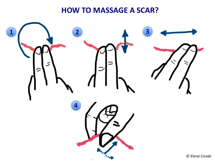
There are different techniques. The most widespread recommendations to facilitate patient autonomy in treatment is to perform cycles with different movements, as shown in the drawing.


We would start with a circular rhythmic movement (Figure 1), with enough pressure to whiten the scar skin but avoiding friction on the epidermis so as not to damage it (we press and move the finger and skin at the same time). We can continue with parallel (Figure 2) and perpendicular (Figure 3) motions. Deep palpation of the scarred skin fold between the thumb and the second finger, called skin rolling, (Figure 4) helps to reduce fibrous adhesions between the skin and deep tissues.

It is widely recommended to perform the massage with a moisturizing product that facilitates the movement of the fingers without damaging the epidermis. Hypoallergenic products should be used to minimize the risk of contact dermatitis.


Do you usually recommend compression therapy for post-surgical or post-traumatic wounds? Do you have any experience with scar massage?


References:

1.    Renò F, Sabbatini M, Lombardi F, Stella M, Pezzuto C, Magliacani G, Cannas M.  In vitro mechanical compression induces apoptosis and regulates cytokines release in hypertrophic scars. Wound Repair Regen. 2003 Sep-Oct;11(5):331-6.

2.    Shin TM, Bordeaux JS. The role of massage in scar management: a literature review. Dermatol Surg. 2012 Mar;38(3):414-23.

3. Cho YS, Jeon JH, Hong A, Yang HT, Yim H, Cho YS, Kim DH, Hur J, Kim JH, Chun W, Lee BC, Seo CH. The effect of burn rehabilitation massage therapy on hypertrophic scar after burn: a randomized controlled trial. Burns. 2014 Dec;40(8):1513-20. 

4. Ault P, Plaza A, Paratz J. Scar massage for hypertrophic burns scarring-A systematic review. Burns. 2018 Feb;44(1):24-38. 

Differences between epidermal and dermo-epidermal punch grafts

Autologous epidermal and dermo-epidermal punch grafts are two of the different options available to promote epithelialization of recalcitrant wounds despite adequate conventional treatment. As we commented in the post “Types of skin grafts to cover chronic wounds: which one should you choose?”, in our wound clinic, the dermo-epidermal punch graft is the technique commonly used. The procedure is outpatient based, simple, economical, and provides a striking analgesic effect on painful wounds. Therefore, it is a technique very well accepted by patients, which can be repeated as many times as necessary to achieve complete epithelialization of the wound. To obtain these dermo-epidermal fragments we can use a punch, scalpel or curette, so their morphology will be more or less circular or oval. The procedure is performed under local anaesthesia in the donor site, usually the thigh , and we do not go deeper than the papillary dermis.

We know that we are at the appropriate depth (papillary dermis) when we find a punctiform bleeding after obtaining each graft. In the image representing the skin layers, blood vessels are distributed vertically in the dermal papillae. If we look at it from above, the vascularization of the papillae would appear as a dotted pattern.

Anatomical knowledge of the skin and experience are key to performing the procedure systematically and quickly. The small grafts are placed directly on the wound bed. Local pressure and immobilization the first few days after the procedure is essential for graft taking. Wounds in the donor site heal by secondary intention. The risk of bleeding and infection of the area from which the grafts are obtained is minimal. One year after the procedure, only slight alterations in the pigmentation (hypo or hyperpigmentation) are observed in the donor site.

Epidermal grafts are obtained by separating the epidemis from the dermis at the dermo-epidermal junction. The epidermis is a flat polystratified epithelium composed mostly of keratinocytes, which are born in the basal layer and progresively differenciate forming the upper layers until their destruction and elimination in the horny layer (the most superficial layer of the skin, which is formed by dead cells).


As the epidermis is not vascularized (its cells are nourished by imbibition through the vessels of the papillary dermis), there will be no bleeding in the donor site and the subsequent scar will be practically imperceptible. In addition, as the dermis remains intact, only the most superficial nerve endings of the dermo-epidermal junction would be stimulated. Consequently, the procedure is practically painless. In order to obtain this type of graft, a device has been marketed which, placed on the skin of the thigh for an average of 30 minutes, produces microvesicles which separate the dermo-epidermal junction. It works by suction using negative pressure and heat, without the need for local anaesthesia. The roofs of these formed blisters are separated by a mechanical cutting system and then are directly applied to the wound.1,2

The main benefits associated with this technique are the ease of use of the marketed device, which can be used in the consultation room without the need for local anaesthesia, and the absence of complications in the donor area by leaving virtually no scar. However, dermo-epidermal punch grafting, despite requiring local anaesthesia and subsequent dressing changes in the donor site (in our clinic we normally use alginate in the first dressing changes), is a technique with low morbidity for the patient and minimal pain and complications in the donor site. When the medical and nursing team have experience with the technique, it may be performed in a short time, resulting highly efficient.

But the differences between the two techniques are not limited to the donor site. Their action in the recipient area is also different. Dermo-epidermal grafts, placed on a wound bed in optimal conditions, achieve wound epithelialization by means of their attachment. However, epidermal grafts would enhance healing by expressing growth factors and promoting the migration of keratinocytes from the edge of the wound. To make an analogy with our observations in our clinic, the behaviour of epidermal grafts may be compared to that of the punch dermo-epidermal grafts when no graft taking is achieved.

An interesting indication for the use of epidermal grafts is the coverage of wounds in the context of pyoderma gangrenosum. Considering that the device only separates the epidermis, we would avoid the phenomenon of patergia in the donor site (a phenomenon that may cause another complicated wound in the area in these patients).3 However, if the activity of pyoderma gangrenosum is controlled with adequate immunosuppressive treatment, other autologous graft options (such as dermo-epidermal punch grafting) may be considered.

Different case series of epidermal grafts have been published (with heterogeneity of aetiologies, extension, evolution time and adjuvant treatments). One of them included 102 wounds, with excellent patient tolerance  and high percentages of epithelialized lesions at the end of follow-up time.4-6 However, a recent systematic review including 7 studies with 209 wounds concludes that high level evidence data is needed to evaluate the real benefit of this type of autologous graft.7 With regard to new studies, a protocol has been designed to perform a multicentre clinical trial to compare the usefulness of dermo-epidermal and epidermal grafts, taking into account the result in the donor and recipient sites.8

Do you have experience with epidermal grafts?


References:

  1. Kirsner RS, Bernstein B, Bhatia A, Lantis J, Le L, Lincoln K, Liu P, Rodgers L, Shaw M, Young D. Clinical experience and best practices using epidermal skin grafts on wounds. Wounds. 2015;27(11):282–92
  2. Herskovitz I, Hughes OB, Macquhae F, Rakosi A, Kirsner R. Epidermal skin grafting. Int Wound J. 2016 Sep;13 Suppl 3:52-6.
  3. Richmond NA, Lamel SA, Braun LR, Vivas AC, Serena T, Kirsner RS. Epidermal grafting using a novel suction blister-harvesting system for the treatment of pyoderma gangrenosum. JAMA Dermatol. 2014;150(9):999–1000.
  4. Hachach-Haram N, Bystrzonowski N, Kanapathy M, Smith O, Harding K, Mosahebi A, Richards T. A prospective, multicentre study on the use of epidermal grafts to optimise outpatient wound management. Int Wound J. 2017 Feb;14(1):241-249.
  5. Everts PA, Warbout M, de Veth D, Cirkel M, Spruijt NE, Buth J. Use of epidermal skin grafts in chronic wounds: a case series. Int Wound J. 2017 Dec;14(6):1213-1218.
  6. Lincoln K, Hyde J. Evaluation of Epidermal Skin Grafts for the Treatment of Complex Wounds in a Wound Care Center: A 94-Patient Case Series. Wounds. 2016 ct;28(10):347-353.
  7. Kanapathy M, Smith OJ, Hachach-Haram N, Bystrzonowski N, Mosahebi A, Richards T. Systematic review and meta-analysis of the efficacy of epidermal grafting for wound healing. Int Wound J. 2017 Dec;14(6):921-928.
  8. Kanapathy M, Hachach-Haram N, Bystrzonowski N, Harding K, Mosahebi A, Richards T. Epidermal grafting versus split-thickness skin grafting for wound healing (EPIGRAAFT): study protocol for a randomised controlled trial. Trials. 2016 May 17;17(1):245.


Types of skin grafts to cover chronic wounds: which one should you choose?

Dibujo injertos 3

The use of skin grafts is a traditional technique to promote the healing of acute and chronic wounds. There are different types of skin graft.

Depending on the origin:

  • Autograft or autologous graft: skin obtained from the patient’s own donor site.
  • Allograft or heterologous graft: skin obtained from another person
  • Xenograft or heterograft: skin from other species, such as pigs.
  • Synthetic skin substitutes: manufactured products that work as skin equivalents. They may be epidermal (keratinocyte cultures), dermal or dermoepidermal (artificial skin).

Only autografts are considered permanent. The rest, either because of their composition (degradable by the host) or because of their origin (rejected by the host’s immune response), only provide temporary coverage during a few weeks.

In our daily clinical practice, in chronic wound clinics, we normally use autologous grafts which, depending on their thickness, are classified into:

  • Total skin graft: includes epidermis and dermis. The incision is made up to the subcutaneous tissue and a wedge of skin is obtained. The edges of the resulting defect in the donor site are approximated by sutures.
  • Split-thickness skin graft: its thickness ranges from 0.2 to 0.4 mm. The cutting plane is located above the hair follicles which, by remaining in the donor site, allow healing by secondary intention. They are divided into thin and thick split-thickness skin grafts.
  • Epidermal graft: it is formed solely by epidermis. The traditional methods for obtaining epidermal grafts, based on the suction technique, were tedious. Recently, a device has been commercialized, Cellu Tome, that obtains epidermal micrografts applying heat and negative pressure on the skin. Separation occurs at the level of the lamina lucida, at the dermoepidermal junction.1 The exact mechanism of action of epidermal micrografts in the wound bed is unknown. Clinical experience seems to indicate that it depends on the quality of the recipient bed. Therefore, in many cases wound reepithelization occurs primarily from the edges of the wound and not by the attachment of the grafts, which would behave as promoters of healing by releasing growth factors.1


Back to the title of this post, which type of graft should you choose? It depends on the wound. As we have seen in the previous post, in order to consider that a wound is prepared for skin grafting, it must meet certain requirements, such as an adequate granulation tissue. However, it is necessary to take into account other factors that will influence the outcome of the intervention, such as the location of the wound, the extent, the underlying pathology and the needs or comorbidities of the patient.

  • When the vascular supply of the wound is low, the use of thin grafts is recommended. This a typical finding in our wound clinic.
  • The contraction of the wound will be greater if there is less viable dermis, such as in thin grafts, which worsens the aesthetics and functionality of the scar. An interesting alternative, used predominantly in extensive wounds and in areas where maintaining mobility is essential, is the use of synthetic dermal substitutes in combination with autologous thin partial thickness skin grafts. Dermal substitutes are sheets with a layer formed mainly by bovine or porcine collagen, which is placed on the wound bed, and another protective silicone layer. After a few weeks, when a suitable dermis is achieved, the silicone sheet is removed and the skin graft is placed.
  • The device to obtain epidermal micrografts (Cellu Tome), might represent an interesting alternative to thin partial skin grafts. This method is simple, does not require anesthesia and minimizes complications in the donor site.1 It is especially interesting in cases of pyoderma gangrenosum because, being minimally invasive, there will be no phenomenon of pathergy in the donor site.2 If you want to know more about epidermal grafts, read the post “Differences between epidermal and dermo-epidermal punch grafts”


What techniques do we normally use to obtain autologous split-thickness skin grafts?

A. Mesh graft.

It is obtained with a dermatome and must be performed in an operating theatre. The main benefit of using a graft with grids is that you can save tissue from the donor site (it can expand several times its size). The interest of this meshing is based on the principle of continuous growth of the keratinocytes, which end up filling the free spaces. On the other hand, this lack of continuity allows fluid to drain through the holes in the graft, which prevents the formation of hematoma or seroma.

For graft taking, it is essential an intimate contact between wound bed and the graft, especially the first 3-5 days. This is not always easily achieved by pressing with gauze and bandages, so the concomitant use of negative pressure therapy may be helpful.


B. Punch grafts (also called micrografts)

Punch grafting does not require the use of complex instruments and can be performed in the consultation room. This is the technique that we normally use in our wound clinic. 

There are different methods of obtaining micrografts, which normally have a diameter between 4 and 6 mm. A curette, scissors, scalpel or punch can be used.3 The fragments obtained from the donor site, which will usually be the anterior face of the thigh, are placed with tweezers on the wound bed, a few millimetres apart from each other. The donor site heals by secondary intention, with the placement of a hydrocolloid dressing or alginate, depending on the bleeding (read post “Which dressing do I choose to cover the skin graft donor site?”). The recipient site is covered with a non-adherent interface dressing and a secondary dressing, or directly with a sheet of calcium alginate fibres as the primary dressing and gauze as the secondary dressing. Local pressure will be applied and compression bandage or discharge will be applied, depending on the aetiology. The first dressing change of the grafted area will be carried out after 5-7 days.


microinjertos
Punch grafting procedure used in our wound clinic. A. Recalcitrant chronic ulcer,without infection data and with adequate granulation tissue. B. Punch grafts on the wound. C. Donor site, thigh, with point bleeding. D. Donor site at day 15. E. Clinical evolution at week 2. Complete epithelialisation at week 6.

Graft taking will depend on the wound bed characteristics (good quality of granulation tissue and little slough), the bacterial load, the immobilization in the days following the intervention and a conservative attitude during the first dressing changes.

Even if not all micrografts adhere to the wound bed, the effect is beneficial in healing, as there is a decrease in pain and growth factors are released. It is essential the conservative cleansing in the first cures, avoiding any kind of debridement in order not to remove the micrografts and maintain the microenvironment that has been created. A few touches with a gauze soaked in physiological serum will be sufficient.


References:

  1. Kirsner RS, Bernstein B, Bhatia A, Lantis J, Le L, Lincoln K, Liu P, Rodgers L, Shaw M, Young D. Clinical Experience and Best Practices Using Epidermal Skin Grafts on Wounds. Wounds. 2015 Nov;27(11):282-92
  1. Richmond NA, Lamel SA, Braun LR, Vivas AC, Serena T, Kirsner RS. Epidermal grafting using a novel suction blister-harvesting system for the treatment of pyoderma gangrenosum. JAMA Dermatol. 2014;150(9):999-1000.
  2. Nordström A, Hansson C. Punch-grafting to enhance healing and to reduce pain in complicated leg and foot ulcers. Acta Derm Venereol. 2008;88(4):389-91.


Punch grafting in postsurgical wounds

This 73 year old patient presents with a scalp wound after removal of a basal cell carcinoma. On the day of surgery, 3 weeks before, wedge resection of the tumor was performed and the edges were approached with a suture to promote healing by secondary intention.

Basal cell carcinoma is the most common cutaneous malignant tumour, followed by squamous cell carcinoma. They usually occur in sun exposed areas and are surgically treated. If direct closure after removal is not possible, the defect can be reconstructed with a flap or graft. Another option is, as in this case, to plan a secondary intention closure. This alternative is common in locations such as scalp or leg, or when part of a flap or graft is necrotic.

In Dermatology Departments we frequently encounter these cases and the question we usually ask ourselves is: how can we speed up the epithelialisation of these wounds? 

In our wound clinic the answer is: with punch grafts (see post: Types of grafts to cover chronic wounds: which one should you choose”, “Which dressing should I choose to cover the skin graft donor site?)

I found inspiration to use this grafting technique beyond chronic leg wounds when I read a poster in the Journées Cicatrisations 2017, in which they presented excellent results of punch grafting on a post-surgical wound on the nasal dorsum and another on the back of the hand.


Here you can see what we did in the case I have presented.

There are different alternatives to cover the grafted site. Alginate calcium dressings directly over the wound is a simple option. If we want to avoid a bulky dressing, there are flexible multi-layer dressings that adapt to complicated anatomical locations for dressing support, such as scalp.

And this is the result in the second post-grafting dressing change, 2 weeks after the procedure.

Obtaining the grafts with a curette in a very superficial way (epidermis and scarce papillary dermis) avoids the cobblestone effect that we may find when using the punch (circular scalpel). In fact, in small wounds with red granulation tissue after necrosis of flaps or grafts on the face, the placement of ultrathin micrografts (with minimal bleeding in the donor area) enhances the release of growth factors and keratinocytes to achieve rapid epithelialization and an excellent cosmetic result (see post: “Differences between epidermal and dermo-epidermal punch grafts”). It is important to remember that dressing changes should be spaced as far apart as possible until complete epithelialization is obtained and, in each dressing change, minimal cleansing should be performed.

We have to bear in mind that a post-surgical wound that takes a long time to close is a stressful factor for the patient. Punch grafting is a simple technique, which may be performed in the consultation room, and which, in addition to promoting epithelialization, reduces pain. Therefore, it is very well accepted by the patient.

I would like to end with a key message: before trying to speed up healing by grafting a post-surgical wound we have to ask ourselves what is going wrong: are there signs of infection, what factors are influencing stagnation? For example, before grafting a recalcitrant post-surgical wound on the leg, we have to consider starting compression therapy to reduce inflammation and enhance healing (See post: “Compression therapy and scar massage in leg post-traumatic or post-surgical wounds). In post-surgical leg wounds, compression therapy and punch grafts are a successful couple in our clinical practice. Here is a case with complete epithelialization in less than a month.

The art of wound cleansing

grifo post 2

It is widely recognized that cleansing is essential in wound management and it is always the first stage in every dressing change. But should you always cleanse the wounds? What is the best way to cleanse the wounds? Saline serum or tap water? What do you normally use? Let’s see what has been studied regarding wound cleansing.


Is wound cleansing always essential?

Wound exudate, especially in acute wounds, is a source of growth factors and other bioactive substances that promote healing. Studies that have compared wound cleansing in acute wounds with its absence, find no significant differences regarding infection and healing rates.1 On the other hand, indiscriminate cleansing may damage the fragile newly formed tissue in the wound bed. Therefore, cleansing any wound should be aimed at eliminating what is hampering its healing, which is not always easy to identify.


Saline serum or tap water?

It stands out the paucity of well-designed studies that have been done to determine the best cleansing solution. The ideal wound cleansing product should be a fluid that helps remove excess exudate and devitalized tissue, without being toxic to the microenvironment of the wound bed. Classical antiseptics (chlorhexidine, povidone iodine) were traditionally used. However, given that their cellular toxicity and consequent slowing of healing have been shown, their use is currently totally discouraged in clinical practice guidelines. This category should not include the new generation of products with antiseptic properties that do not alter the healing process, such as polyhexanide, whose use is beneficial in recalcitrant wounds with resistant biofilm and signs of local infection.

A systematic review has recently concluded that drinking tap water may be as effective and more efficient (because of its lower cost) than sterile water or saline serum. Seven clinical trials comparing infection and  healing rates when comparing the use of tap water and saline serum  were included and no statistically significant differences were detected in both acute and chronic wounds.1


What are the properties of each solution?

0.9% saline serum is an isotonic solution. Consequently, it does not alter wound bed fluids, does not hinder healing, does not cause tissue damage, produces no allergy and does not alter normal bacterial flora.

Water has been used for years, and its benefit has been proven. Water is a hypotonic solution, so long-length or frequent water irrigation would stimulate the absorption of fluid by osmosis, with the consequent increase in oedema and cell rupture. This would increase exudate and, consequently, the need of dressing changes. However, while cleansing does not involve immersion of the wound in water for a prolonged period of time, its regular use in wound cleansing is not only safe, but also easily accessible and efficient.2 In countries with limited resources and difficult access to drinking water, an alternative is the use of boiled or distilled water.

Another very interesting point: What is the optimal temperature at which the cleansing solution should be?

The optimal body surface temperature for the healing process is in the range of 33 to 42ºC. Outside these limits, healing is delayed.

During each dressing change, wound bed temperature decreases, inhibiting mitotic activity. Recovery from normal temperature may take 40 minutes, while restart of mitosis may occur up to 3 hours later.

It has been shown that cleansing solutions used at room temperature can lower wound bed temperature by 2°C, and therefore wound bed temperature would be less than 33°C. Consequently, in practice, it would be ideal to heat water or saline serum to at least 37°C. Moreover, cleansing should be rapid to avoid a drop in temperature.3


Referencias:

  1. Fernandez R, Griffiths R. Water for wound cleansing. Cochrane Database Syst Rev. 2012 Feb 15;2:CD003861. doi: 10.1002/14651858.CD003861.
  1. Flanagan M, Fletcher J. Wound care: the healing process. Nurs Stand. 1997 Jun 25;11(40 Suppl Nu):5-17.
  1. McGuiness W, Vella E, Harrison D. Influence of dressing changes on wound temperature. J Wound Care. 2004 Oct;13(9):383-5.


Silver in wound healing

Why?

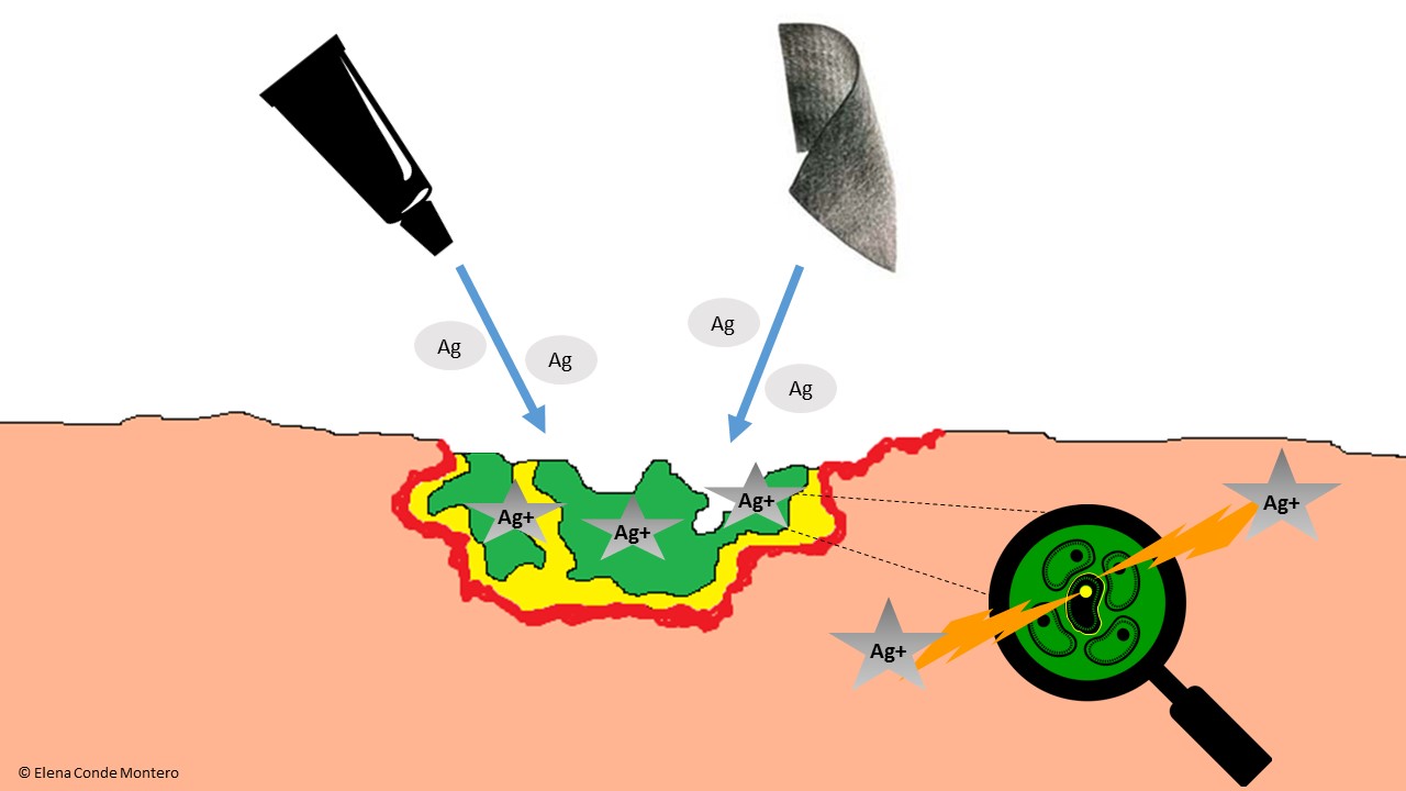
The antimicrobial activity of silver has been demonstrated in vitro, including multi-resistant bacteria such as MRSA. To be bactericidal, silver has to turn into silver ion (Ag+). Wound exudate favours this transformation, either by loss of an electron if silver was in its elemental form, or by separation of the compound to which the silver ion was attached.

Silver ions are very reactive and produce bacterial destruction by their action in different targets: they break down the bacterial wall, interfere with protein production and cell replication. Different studies in experimental models suggest a reduction of bacterial adherence and destabilization of the biofilm matrix (See figure).

mecanismo accion plata VF

One well-designed clinical trial and two Cochrane reviews conclude that the use of silver dressings may not improve the healing process,1-3 which contrasts with the results of other studies, expert opinions and the observations in our clinical practice. These results may be due to inappropriate use of silver dressings, in wounds without signs of infection or for long periods of time. I would like to highlight a review published in the journal of the American Academy of Dermatology4 that supports the beneficial effect of silver in decreasing wound size, without being able to determine its role in long-term healing rates.


How?

Silver may be present in the dressing structure or as a coating and can be found in different forms:

  • Elemental silver: nanocrystalline silver.
  • As an inorganic compound: silver oxide, silver phosphate, silver chloride, silver sulfate, silver sulfadiazine.
  • As an organic compound: silver alginate, ionic silver-impregnated sodium carboxymethylcellulose.

Different dressings have variable amounts of silver and, when comparing their in vitro antibacterial efficacy, no correlation has been found between the content or amount of silver released and their antimicrobial activity.

Silver in the dressing needs to contact wound exudate so that it can turn into its ionic form and can perform its antibacterial action. Therefore, although silver content is important, many other factors influence the ability of a dressing to destroy microorganisms, such as the distribution and availability of silver in the dressing and the appropriate choice of the dressing depending on its adaptability and absorption capacity according to wound exudate.5


When?

Silver use in wound healing would be consensually indicated in two situations:

  • Therapeutic indication, in ulcers with excessive bacterial load or signs of infection.
  • Prophylactic indication, in wounds at risk of infection.

The limit of silver use is not well defined. It has been proposed that an evaluation should be performed after 2 weeks to decide whether to continue its application:5

  • If the wound has improved, but signs of infection persist, the silver dressing would be continued.
  • If there is no clinical improvement, its use should be discontinued.
  • If the evolution is good and there are no clinical signs of infection, it should also be discontinued.

The risk of systemic toxicity from prolonged use of silver dressings has been suggested. Generalized argyria is due to the deposition of silver-containing particles in different organs. The systemic toxicity of silver is very low, and its effects are limited to cutaneous-mucosal hyperpigmentation. The minimum amount of silver needed to produce it is not known, but it appears that it could be triggered with oral doses greater than 3.8 g of elemental silver. The amount of silver systemically absorbed in its use in wounds is minimal.6 Although a local greyish pigmentation can be produced by silver deposit, it should not be confused with a generalized argyria.

If you want to know more about silver in wound healing: Appropriate use of silver dressings in wounds: international consensus document

This is a consensus document following a meeting of experts in 2011 to formulate internationally recognised guidelines on the appropriate use of silver dressings, based on clinical practice experience and available evidence.


References:

  1. Michaels JA, Campbell B, King B, et al. Randomized controlled trial and cost-effectiveness analysis of silver-donating antimicrobial dressings for venous leg ulcers (VULCAN trial). Br J Surg 2009; 96(10): 1147-56.
  2. Vermeulen H, van Hattem JM, Storm-Versloot MN, Ubbink DT. Topical silver for treating infected wounds. Cochrane Database Syst Rev 2007; 24(1): CD005486.
  3. Storm-Versloot MN, Vos CG, Ubbink DT, Vermeulen H. Topical silver for preventing wound infection. Cochrane Database Syst Rev 2010; 17(3): CD006478.
  4. Carter MJ, Tingley Kelley K, Warriner RA. Silver treatments and silver impregnated dressings for the healing of leg wounds and ulcers: A systematic review and meta-analysis. J Am Acad Dermatol 2010; 63: 668-79.
  5. Consenso Internacional. Uso adecuado de los apósitos de plata en las heridas. Consenso del grupo de trabajo de expertos. London: Wounds International, 2012.
  6. M C Fariña, P Escalonilla, R Grilli, M L Soriano, L Martín, L Requena, J L Sarasa, A de Castro. Argiria generalizada secundaria a la administración tópica de nitrato de plata. Actas Dermosifiliogr. 1998;89:547-52.


Band and bandage: not the same thing

A band is called a bandage when it is placed on the patient’s leg and, therefore, its properties are different. A band may be more or less elastic, while a bandage may have more or less rigidity. Bearing in mind that we have just celebrated the II European Conference on Venous Ulcer and Compression, in which it has become clear that WITHOUT TRAINING THERE IS NO OPTION! we are going to remember basic concepts to understand compressive therapy. Before continuing, I invite you to visit the Educational tools section of the website of the International Compression Club, where you will find more details of the concepts that we are going to discuss. Let’s start the training!


Extensibility and elasticity

These are two in vitro properties of a band.

Extensibility is the ability of a band to stretch in response to an applied tension.

Elasticity is the ability to return to its original length when that tension is reduced.

Simply put, a band with great extensibility will also have great elasticity and vice versa. To classify bands according to these properties, the terminology short-stretch and long-stretch is used in Europe. In Spain, professionals usually talk about low and high elasticity bands (also called inelastic and elastic).

In a reductionist way, we commonly speak of elastic bandages to refer to those that make pressure during both rest and exercise, adapting to the changing diameter of the calf, while we consider inelastic bandages when their pressure at rest is low and increases in peaks with muscle contraction. If we place a single band, elastic or inelastic, its in vitro properties may approach those in vivo. However, as we will see below, a bandage is more complex than a band.


Multilayer and multicomponent

The first term is an inherent feature of any bandage. Any bandage placed on one leg will be a multi-layer bandage, even if only one bandage is applied, as there will always be some overlap between layers.

If one or several single-type bands are applied, the bandage will be single-component. If we overlap bands with different physical properties, the bandage will be called multicomponent.


Pressure under the bandage

For healing promotion of venous ulcers, if no contraindication exists, the consensual recommendation is to apply a strong pressure on the ankle (>30-40 mm Hg). This measure is taken with a device that is placed on the ankle and indicates the pressure exerted at that point during rest. However, the pressure exerted is different at each point of the leg and depends on different factors, all of which are included in the famous Laplace´s Law:

The pressure under the bandage at a given point is higher the greater the tension (force applied to the bandage during placement), the greater the number of layers, the smaller the radius of circumference at that point of the limb (greater prominence, e.g. pretibial region) and the narrower the width of the bandage.

The ability to maintain over time, and during movement, that degree of pressure depends on the properties of the bandage components.

Did you know that all the padding techniques we use in our clinical practice have their explanation in this physical law? Let’s analyse them: 

  • Padding to homogenize the perimeters of the leg in patients with alterations in the morphology of the limb. The objective is to obtain an adequate pressure gradient, as in legs with physiological proportions. The bandage will exert a decreasing pressure from the ankle to the calf, as the diameter of the limb increases and therefore exerts less pressure in the proximal area.


  • Padding to protect bony prominences and other sensitive areas from hyper pressure. The pretibial region or the Achilles tendon area are two locations that require special protection to avoid damage from local excessive pressure.
  • Padding to increase retromaleolar pressure. Compression therapy to promote healing or prevent the recurrence of wounds located behind the malleolus must overcome the “tent” effect that usually occurs in that concave area after placement of the bandage or compression stocking. The use of padded devices to fill this gap (always with bevelled edges and placed with the smaller surface side on the skin) helps to increase local pressure and therefore the effectiveness of compression therapy.

Stiffness

It is a property of the bandage, that is to say, an in vivo property. It is defined as the resistance of the bandage to changes in calf muscle volume during movementThe greater the stiffness, the greater the effectiveness in reducing venous hypertension while walking (ambulatory venous hypertension). By presenting resistance to muscular contraction, high pressure peaks are achieved and, consequently, brief and intermittent venous occlusions, similar to physiological valvular function, occur. At rest, these peaks do not occur and the pressure is lower. This difference in pressure is called the Dynamic Stiffness Index, which is greater the more resistance the bandage presents to muscular contraction (to put it another way, “greater impact of the muscle against the bandage”).

If the bandage is not very rigid, there are no pressure peaks during activity, as it adapts to changes in calf shape, with little resistance to increased muscle volume. The pressure under the bandage in these cases does not vary much between rest and activity (lower Dynamic Stiffness Index).

The concept of more or less rigid bandage should replace the current classifications of “inelastic and elastic bandages”. As we have seen, elasticity is a property of the bandage outside the patient. In single-component bandages the simplification “inelastic bandage = inelastic bandage” and “elastic bandage = inelastic bandage” can be practical in our daily life. However, taking into account that all bandages are multi-layer and many are multi-component, the mechanical characteristics of the bandage vary with respect to those of the bands that compose it. In fact, friction between the layers of a bandage, especially if there is a cohesive component, increases the stiffness of the bandage. Likewise, if we apply an elastic band on a herringbone pattern, we get a more rigid bandage.

And what is the cutting point to differentiate the bandages according to their greater or lesser stiffness?

The differentiation between more or less rigid bandages is made using the Static Stiffness Index (SSI). This index is the difference in pressure under the bandage that occurs when the patient moves from lying down to standing. If the pressure difference is greater than 10 mmHg, it is considered a high stiffness bandage and if it is lower, a low stiffness bandage.

All this helps us to understand the reason why muscular atrophy or mobility limitation decreases the effectiveness of compressive bandages. But this immobility should not be the reason for not applying compressive therapy, nor should a certain degree of arteriopathy be. In immobile patients with peripheral artery disease with ITB>0.5, bandages with a high stiffness index and pressure below 30 mmHg is a safe and effective option if foot dorsiflexion exercises are performed.

This is because muscle contraction during dorsiflexion movements of the foot produces pressure peaks that facilitate venous return, with the consequent decrease in venous hypertension. In addition, we can have a double benefit, since the resulting reduction in oedema can improve arterial flow.